Tuesday, 1 November 2005 - 2:22 PM
233h
Evaluation of Polymer and Self-Assembled Monolayer Coated Silicone Surfaces to Reduce Neural Cell Growth
Kruti R. Patel1, Haiying Tang2, William E. Grever3, K. Y. Simon Ng2, Jianming Xiang4, Richard F. Keep4, Ting Cao2, and James P. McAllister II5. (1) Department of Biomedical Engineering and Department of Pediatric Neurosurgery, Wayne State University, 4201 Antoine Street, UHC-6E, Detroit, MI 48201, (2) Department of Chemical Engineering, Wayne State University, 5050 Anthony Wayne Drive, Detroit, MI 48202, (3) Children's Research Center of Michigan, Department of Pediatrics,School of Medicine, Wayne State University, 3L35, Detroit, MI 48201, (4) Department of Neurosurgery, University of Michigan, 5550 Kresge I, Ann Arbor, MI 48109, (5) Department of Pediatric Neurosurgery,Children’s Hospital of Michigan, Wayne State University, 4201 Antoine Street, UHC-6E, Detroit, MI 48201
Chemical vapor deposition method was used for coating FAS on silicone. OTS was coated on silicone after plasma treatment. Heparin and hyaluronan were coated with photo-immobilization method on OTS modified silicone surfaces. Contact angle measurements confirmed that the hydrophobic and hydrophilic properties of these coatings were stable on the silicone surfaces for at least 30 days, indicating that coatings were stable during cell culture (approximately 14 days). Choroid plexus cells and astrocytes were obtained from the brains of 1-2 day old Sprague-Dawley rat pups. The enriched choroid plexus cells and astrocytes were cultured for two weeks and one week respectively on the unmodified and modified silicone samples secured with teflon rings in polystyrene 12-well plates. Since the silicone samples were opaque and visual inspection of cell growth on silicone samples was difficult, the polystyrene 12-well plate surface was used as a control to determine the stage of confluence for cell cultures. Cells were counted using a hemocytometer, which provides an indirect measure of cell growth and adhesion, since attachment to a substrate is a requirement for prolonged survival in culture.
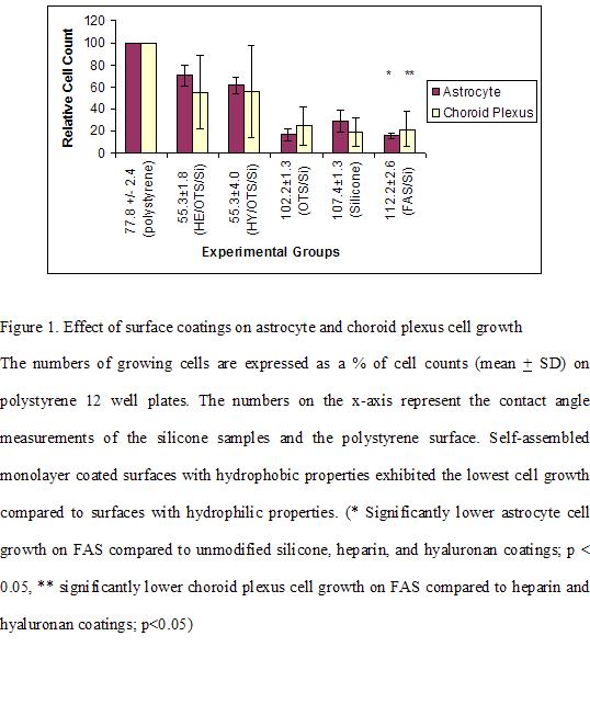
Compared to unmodified silicone, FAS (p<0.05) coated surfaces significantly reduced astrocyte proliferation, while no significant difference in astrocyte growth was observed on OTS (Figure 1). Heparin (p<0.001) and hyaluronan (p<0.001) coated surfaces resulted in an increase in astrocyte growth in comparison to unmodified silicone surfaces. A similar trend was observed for choroid plexus cell growth on heparin (p<0.05) and hyaluronan (p<0.05) coatings compared to unmodified silicone surfaces. However, no significant reduction in choroid plexus cell proliferation was observed on FAS or OTS coated surfaces compared to unmodified silicone surfaces. Low cell growth may be attributed to hydrophobicity of the surfaces (FAS 112.2±2.6 0, OTS 102.2±1.3 0). Furthermore, atomic force microscopy measurements revealed that unmodified silicone had the roughest surface while coatings decreased the surface roughness of silicone. However, surface roughness did not play an important role on cell growth. Unmodified silicone with the roughest surface (RMS 153.8 ± 46.1) exhibited low cell growth compared to heparin and hyaluronan with surface roughness of 104.0 ± 20.7 and 96.8 ± 18.2 respectively. FAS (RMS 81.1 ± 26.4) and OTS (RMS 88.8 ± 12.5) coatings had comparatively smoother surfaces but the lowest cell growth of all the samples. The results of this study indicate that silicone shunts coated with the self-assembled monolayer (FAS) may be suitable for future clinical applications to improve the treatment of hydrocephalus.
See more of #233 - Polymers for Biofunctional Surfaces (08A14)
See more of Materials Engineering and Sciences Division
See more of The 2005 Annual Meeting (Cincinnati, OH)