Tuesday, November 6, 2007 - 2:10 PM
212e
Dielectrophoretic Manipulation of Mycobacterium: Toward High Throughput, Post-Mutagenesis Screening for Membrane Lipid Mutations
Introduction This presentation reports the state of development of a dielectrophoresis-based, high-throughput, post mutagenesis screening technique for membrane lipid mutations in Mycobacterium. We have successfully actuated Mycobacterium smegmatis using continuous-flow, insulative dielectrophoresis; controlling the transverse position of an input stream of cells in glucose media by varying the applied electric field magnitude. Mycobacterial infectious diseases include leprosy, tuberculosis (TB), and Mycobacterium avium complex (MAC). While incidence in developed nations is low, tubercular infections and fatalities continue to increase worldwide, affecting a significant portion of third-world and developing nations1. MAC infections kill a continually increasing number of AIDS and other immuno-compromised patients worldwide1. In addition to rising rates of infection, the emergence of multi-drug resistant (MDR) strains of both TB and MAC makes the study of these diseases imperative. Mycobacterial infections are characterized by the formation of encapsulating granulomas – macrophage-encapsulated Mycobacterium bacilli surrounded by inflamed tissue – caused by the glycolipids that make up the Mycobacterium cell envelope. The mechanisms by which Mycobacterial cell wall lipids generate this response is not well understood2. While the genome of M. tuberculosis is known, the precise function of membrane lipids and their role in the Mycobacterium-host cell interactions is an active area of research3. This, as well as the emergence of MDR strains of Mycobacterium, illuminates a critical gap in the current toolset used by researchers developing pharmaceuticals and treatment techniques: no library of membrane lipid mutations currently exists for Mycobacterium. Mycobacterium tuberculosis, in particular, is difficult to culture in vitro and has a long cell cycle time, making development of such a library problematic. Random transposon mutagenesis is one potential technique that can generate a large number of unknown mutations, but no technique exists for identifying those with membrane specific differences in a rapid, high-throughput device, and the time and cost associated with characterizing and sequencing all mutations produced is prohibitive. We present a technique that fills the gap between the generation of phenotyped populations and a library of lipid mutations in Mycobacterium, by interrogating numerous cells simultaneously based on membrane characteristics.
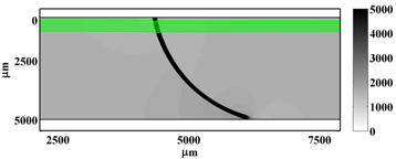
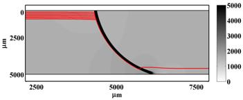
Mycobacterial envelope properties influence the cell's effective electrical permittivity and conductivity and therefore change a cell's electrical response as a function of membrane composition. We take advantage of this change in electrical character using dielectrophoresis (DEP). Dielectrophoresis – particle motion in response to a non-linear electric field – is induced using insulating constrictions in the depth of a microchannel. The DEP force is proportional to the gradient of the squared electric field, with a constant of proportionality that is particle-dependent. Constrictions in channel depth increase the local electric field, causing gradients that induce a local dielectrophoretic force normal to the constriction. In addition to non-linear dielectrophoresis, the DC-offset, AC electric field applied in this device (at reservoirs located at microchannel outlets) generates linear electroosmotic fluid flow and linear electrophoretic particle motion everywhere in the device. We present novel, 3D, modulation of electric fields in microfluidic devices . Most insulative DEP devices trap particles by countering bulk particle motion (the result of electroomosis, electrophoresis, and/or pressure-driven flow) with local, dielectrophoretic forces, creating a point where the net particle velocity is zero. Insulating constrictions in the width of a microfluidic channel are traditionally used to generate local DEP forces. We, however, constrict the depth of a microchannel, introducing an additional degree of freedom: the angle of incidence between bulk linear electrokinetic forces and opposing nonlinear DEP forces (Figure 1). We then vary the angle of incidence continuously across the channel width, transverse to the direction of flow, by using a curved constriction in channel depth, creating a DEP force that decreases with transverse position. Particle paths in the presence of this curved constriction are a function of the relative magnitudes of electrophoretic, electroosmotic and dielectrophoretic forces. Particle motion far from the constriction is governed by linear electrokinetic forces. As a particle approaches the constriction, DEP forces are generated perpendicular to the constriction. Depending on particle dependent nonlinear DEP forces, the particle will either pass the ridge or be deflected tangent to the constriction. Particles deflected tangent to the constriction will experience a decreasing DEP force, due to variation in the angle of incidence, and eventually pass over the ridge when linear electrokinetic forces dominate, at a point determined by particle electrical characteristics (Figure 2). DEP forces on a particle near a constriction are a function of particle properties and constriction angle of incidence. Whether particles pass through or are deflected tangent to the constriction is determined by the ratio of linear electrokinetic forces to non-linear DEP forces, i.e., is a function of particle or cell properties.
| |
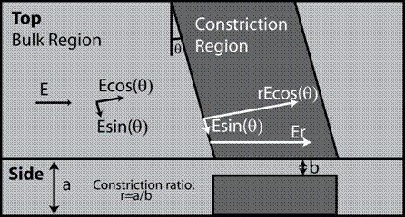 Figure 1. Constrictions in channel depth create electric field gradients that drive DEP forces normal to the constriction. The magnitude of the gradient is therefore dependent on the constriction ratio – bulk channel depth to constriction region depth – and the ridge angle, q. Combined AC and DC electric fields, in the absence of pressure-driven flow, independently modulate linear (electroosmotic and electrophoretic) and non-linear (dielectrophoretic) particle velocity components. We can therefore control linear electrokinetic phenomena by changing the applied DC electric field and non-linear electrokinetic phenomena by controlling the ratio of AC to DC electric fields. |
Figure 2. Top view of device, showing numeric solutions for 3D electric fields in the presence of curvilinear constrictions in channel depth, combined with a mobility model for particle motion, allow for rapid, model driven device design for specific applications. The solutions above, for a curvilinear constriction in channel depth, predict particle deflection transverse to the direction of flow as a function of DEP mobility. |
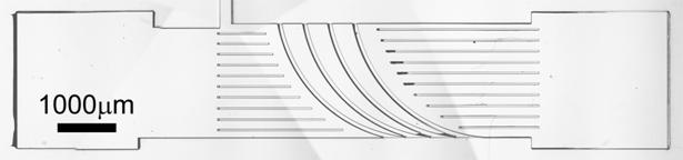
Figure 3. Top view of device. Microchannels are fabricated in Zeonor polymer with curvilinear constrictions in channel depth that create spatial variations in the channel cross sectional area and the gradients responsible for DEP. Dielectrophoretic trapping effects are modulated by continuous variation of the angle of incidence, q, transverse to the direction of flow. Coherent patterning of curvilinear constriction structures causes additive particle transport and increased separation resolution. The resulting output stream has a continuous variation of particle electrokinetic properties transverse to the direction of flow. Device is 1cm in total length. | |
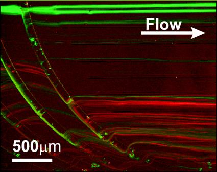
Figure 4. Top view of device showing continuous flow separation of 1.7 and 2.0 micron diameter polystyrene spheres using localized DEP forces to deflect an input stream of particles – image is a time average of 30 frames. Flow direction is from left to right and channel width is 2.5mm. |
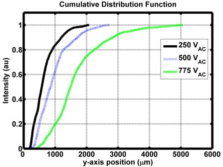
Figure 5. Fluorescently labeled M. smegmatis cells show a variation in output distribution as a function of applied AC electric field. Increased deflection transverse to flow is a characteristic of increased DEP force applied to particles in the presence of the curved constriction. |
(1) World Health Organization; World Health Organization, 2006; Vol. 2007. (2) Geisel, R. E., Sakamoto, K., Russell, D.G., Rhoades, E. R. Tuberculosis 2005, 85, 159-176. (3) Elizabeth R. Rhoades, R. E. G., Barbara A. Butcher, Sean McDonough, David G. Russell Tuberculosis 2005, 85, 159-176. (4) Benjamin G. Hawkins, A. E. S., Yusef A. Syed, Brian J. Kirby Analytical Chemistry 2007 Submitted. (5) Wallow, T. I.; Morales, A. M.; Simmons, B. A.; Hunter, M. C.; Krafcik, K. L.; Domeier, L. A.; Sickafoose, S. M.; Patel, K. D.; Gardea., A. Lab on a Chip 2005, Accepted. | |
Web Page: www.kirbyresearch.com/