Thursday, November 8, 2007 - 10:40 AM
522g
Nano- And Micro- Scale Replication Of Intestinal Basement Membrane Using Chemical Vapor Deposition
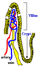
The oral drug delivery market is the largest branch of the total drug delivery market. Over the next several years it is expected to grow at a compound annual growth rate of 10%, reaching upwards of $52 billion by 2010 . An important step in development of oral drug compounds is testing their ability to pass the intestinal epithelium. These tests are typically done with cell culture systems that unfortunately do not, in many cases, accurately predict oral drug absorption. It is hypothesized that, in the study of oral drug transport, cells cultured on a biomimetic substrate model will more accurately reflect in vivo cellular behavior as compared to those cultured on a flat substrate. Specifically, it is expected that biomimetic nano- and micro-topology will influence expression of proteins important to drug uptake and metabolism, because these proteins are expressed differentially along the villus-crypt axis of the intestinal basement membrane in vivo. Fabricating a biomimetic mold of a section of the small intestine for culture of small intestinal epithelial cells will enable testing of this hypothesis. Pig intestine, believed to be similar to human intestine, is being used as the template for the biomimetic scaffold. The pig intestine is cleaned of contents and treated to remove mucus and epithelial cells, so that intestinal basement membrane will provide the micro- and nano-topology that will be replicated. The removal of cells is accomplished by first fixing the tissue in glutaraldehyde and paraformaldehyde and then macerating cells with 0.1% Osmium tetroxide in PBS. In order for intestinal basement membrane to be able to be used in a chemical vapor deposition (CVD) process, it must be able to withstand a vacuum. It is thus prepared in a manner similar to that used for SEM preparation, since the vacuum utilized in a typical SEM is much greater than that used in the silica deposition CVD process. The samples are dehydrated and critical point dried. SEM and histology are used to verify that all cells have been removed and the basement membrane topographical features are intact. The topology of the intestinal basement membrane is being replicated using a chemical vapor deposition (CVD) technique to create a thin film of silica on a section of intestinal basement membrane prepared as described above. A design of experiments (DOE) was carried out to determine the optimal reaction conditions. It was discovered that running at a reactant ratio of between 1:5 and 1:10 methyl silane: hydrogen peroxide,a pressure of 3000 mtorr, and a stage temperature of 5° C produced the desired thin film of silica. At 5° C, a silica film was produced at a deposition rate of 10 nm/min. This process is currently being optimized to coat a fixed section of intestinal basement membrane with silica to a thickness of 1 micron. Once the silica mold has been fabricated, the biological template will be removed by exposure to a strong basic solution, leaving the free-standing scaffold. CVD will again be used to deposit a biodegradable polymer membrane onto the silica mold. This polymer membrane will be peeled off of the mold and used for culturing intestinal epithelial cells. The polymer proposed for the membrane is Poly(2-hydroxyethyl methacrylate), otherwise known as pHEMA. pHEMA has been widely used in cell culture because it is biocompatible and its porosity can be controlled by adjusting the degree of crosslinking, making it ideal for drug transport studies. To aid in development of process conditions, pHEMA was deposited on a flat surface using plasma CVD. Ratios of 5:4:1 of Argon, monomer 2-hydroxyethyl methacrylate (HEMA), and tert-butyl peroxide (TBPO), respectively, were used. Argon served as an inert gas to keep the flow in the reactor constant, and TBPO was added as the initiator. pHEMA was produced running a plasma at 20 Watts. To use pHEMA as a culturable substrate, crosslinking of pHEMA using ethylene glycol diacrylate (EGDA) will be necessary to lengthen the degradation time. Experiments were run to determine deposition rate of the pHEMA alone and EGDA alone to find the best crosslinking ratio. Ongoing studies include crosslinking of the polymer at different ratios to provide samples for a degradation rate study. In summary, results to date demonstrate the ability to fabricate thin films of silica and pHEMA, which will be fabricated with biomimetic topography and utilized for cell culture in future studies.
See more of #522 - Biomimetic Materials I (08B16)
See more of Materials Engineering and Sciences Division
See more of The 2007 Annual Meeting
See more of Materials Engineering and Sciences Division
See more of The 2007 Annual Meeting