
We are investigating the effect of solid/liquid interfaces on the formation of amyloid fibrils by tailored polypeptides. The solid surfaces that we have used are a series of liposomes that include charged and neutral lipids and two which resemble mammalian cells membranes. Six different types of liposomes were prepared: a) 4:1 (phosphatidylcholine, PC: phosphatidylserine, PS) b) 4:1 (cholesterol:PC); c) 1:4 (cholesterol:PC), d) 2:2:1:1 (cholesterol:PC:phosphatidylglycerol, PG: phosphatidylethanolamine, PE), this liposome resembles the myelin membrane, e) 10:5:7.5:16 (PC:PE:PS:cholesterol), this liposome mimics rat's neural membranes and f) dieleoylphosphatidylcholine/dioleoylphosphatidylglycerol
We have characterized these liposomes by measuring (osmotic) second virial coefficients at various pHs and temperatures. This has been done in an attempt to correlate potentials of mean force with the induction (or inhibition) by these liposomes of fibril formation.
Static light scattering experiments were performed using a Brookhaven temperature controlled goniometer equipped with a 2W argon ion laser. The data was analyzed using proprietary software (Brookhaven) to obtain second virial coefficients. The change in refractive index with liposome concentration was measured with a differential refractometer (Brookhaven).
Liposomes were prepared as follows. 20 mg of the lipids were dissolved in 1 ml of tert-butylalcohol. Sucrose (7.5 fold the amount of lipids) was dissolved in 1 ml of water. The solutions were thoroughly mixed and sonicated for 30 seconds. If the lipid did not dissolve in 1 ml of tert-butylalcohol up to 1 addition ml of the alcohol was added. After mixing, the solution must be optically clear. The mixture was filtered through a 0.22 μm filter. The sample was freeze dried for up to 8 hours. Liposomes were produced by adding from 2 to 4 ml of the appropriate buffer to 20 mg total lipids. This was followed by sonication and extrusion through a 0.2 μm filter. After that the solution was diluted up to 1000x to determine the size of the preparation by dynamic light scattering (using the same instrument)
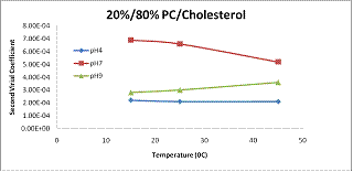
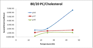 The
figures below show some representative data for two of the preparations. These
two liposom preparations are almost identical size (as determined by dynamic
light scattering) but one is more rigid than the other. There are striking
differences in the behavior of the second virial coefficients. The liposomes
with a higher content of PC have second virial coefficients that are one order
of magnitude larger than the liposomes with a low PC content. Moreover, the
temperature seems to have the opposite effect on the second virial coefficient
of both preparations. We are currently calculating potentials of mean force to
rationalize these findings.
See more of this Group/Topical: Engineering Sciences and Fundamentals