
 Google
 Yelp
 Facebook
 Twitter
 ChEnected
268772 Dielectrophoretic Isolation of DNA and Nanoparticles From Whole Blood
268772 Dielectrophoretic Isolation of DNA and Nanoparticles From Whole Blood
Monday, October 29, 2012: 10:15 AM
408 (Convention Center )
AIChE 2012
Topical 3: 2012Annual Meeting of the American Electrophoresis Society “T3006 Advances in Electrokinetic and Electrophoresis: Fundamentals” Dielectrophoretic Isolation of DNA and Nanoparticles Directly from Whole Blood Michael J. Heller1,2, Avery Sonneberg2, and Raj Krishnan3University of California San Diego, Departments of Nanoengineering1 and Bioengineering2, Atkinson Hall Rm 2312, La Jolla, CA, 92093-0448, and Biological Dynamics3, 9393 Towne Centre Dr., San Diego, CA 92121 Abstract - Dielectrophoretic (DEP) devices and methods have been developed which allow the rapid isolation, concentration and detection of high molecular weight (hmw) DNA and nanoparticles directly from whole blood. Using special microarray devices operated at 20 volts peak-to-peak and 10 kHz, a wide range of fluorescent hmw-DNA and fluorescent nanoparticles can be concentrated into DEP high-field regions while blood cells are concentrated into the DEP low-field regions. A simple fluidic wash then removes the blood cells while the DNA and nanoparticles remain concentrated in the DEP high-field regions where they can be detected by fluorescence. Hmw-DNA could be detected at 260 ng/ml, which is a detection level suitable for cell free circulating (cfc) DNA biomarker analysis. Fluorescent 40 nm nanoparticles could be detected at 9.5 x 10⁹ particles/ml, which is a level suitable for monitoring drug delivery nanoparticles. These DEP devices are now also being used to isolate cfc-DNA from Chronic Lymphocytic Leukemia (CLL) and other cancer patient whole blood samples. Blood sample to PCR is achieved in less than twenty minutes. The ability to rapidly isolated and detect disease related DNA biomarkers and nanoparticles from undiluted whole blood will benefit many diagnostic applications by significantly reducing sample preparation time, complexity and costIntroduction - AC electrokinetic methods like dielectrophoresis (DEP) are well-known techniques for achieving effective separations of cells, nanoparticles, and biomolecules. Until recently, DEP technology had remained impractical for general use with complex biological samples (undiluted blood) and in high conductance solutions (>0.1 S/m). For example, earlier DEP work on separating bacteria from blood (~7-9 mS/cm) required a 50-fold dilution of the blood sample before the DEP separation process could be carried out. Many other DEP separations, including cells, virus, polystyrene nanoparticles, DNA and proteins also required low-conductance conditions. More recently, some progress has been made by several groups on carrying out DEP under high conductance conditions. We for one, have developed new DEP devices and methods that allow both hmw-DNA and nanoparticles to be isolated and detected in high ionic strength buffers (Krishnan R., Sullivan B.D., Mifflin R.L., Esener S.C., & Heller M.J., AC electrokinetic separation & detection of DNA nanoparticles in high-conductance solutions, Electrophoresis 29, 1765-1774, 2008; Krishnan R. & Heller M.J., AC electrokinetic method for enhanced detection of DNA nanoparticles, J. Biophotonics 2, 253-261, 2009; Krishnan R., Dehlinger D.A., Gemmen G.J., Mifflin R.L., Esener S.C., & Heller M.J., Interaction of nanoparticles at the DEP microelectrode interface, Electrochem. Comm. 11, 1661-1666, 2009). Using these special microarrays with robust platinum microelectrodes over-coated with a thin hydrogel layer, DEP separations in high conductance buffers, and now whole blood, can be carried out where hmw-DNA and nanoparticles are concentrated into the DEP high-field regions over the microelectrodes, while micron size particles and blood cells are concentrated into the DEP low-field regions between the microelectrodes. Other groups have used lateral flow driven continuous DEP microseparators to separate blood cells in high conductivity media, and hybrid electrokinetic techniques (DEP, electrophoresis, electrothermal flow) to separate bacteria from blood cells under high conductance conditions. Nevertheless, our results appear to be the first that show the isolation and concentration of hmw-DNA and nanoparticles into well defined high-field regions on the microarrays is due mainly to DEP. Results: Figure 1 (below) shows the original electric field microarray device used for the initial DEP separation work. The expanded view shows how the DEP high-field (red) regions and DEP low-field regions (green) are created when and AC electric field is applied to nine microelectrodes. Original DC/AC electric field microarray devices were obtained from Nanogen and have one hundred 80um platinum microelectrodes overcoated with a thin hydrogel layer. Newer dielectrophoretic (DEP) microarray devices specifically designed for the isolation of cfc-DNA from high conductance buffer and blood samples were obtained from Biological Dynamics. In general, DEP experiments were carried out by adding about 20 ul of blood sample to the DEP microarray device. The DEP field was applied at 10 kHz and 20 Vp-p for 15 minutes to the nine microelectrodes. The microarray was then washed three times with 0.5x PBS with the DEP field on, and finally examined by epifluorescent microscopy.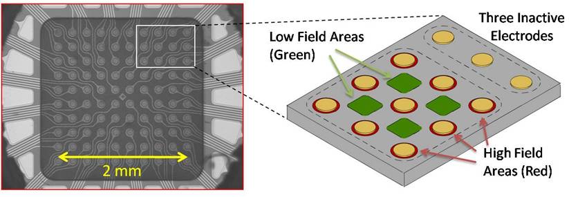 Figure 1 - DEP electric field microarray device and the DEP high-field and low-field regions Figure 2 (below) shows the results for the DEP separation of single-stranded (ss) and double-stranded (ds) high molecular weight (hmw) DNA from whole blood and a whole blood (no DNA) control (6 to 8 mS/cm). All images are in green fluorescence. The DEP AC voltage was applied to the nine electrodes in columns 2, 3, 4, while no voltage was applied to the three electrodes in column 1, which serve as negative controls. (a) Shows the results for a 20 µl sample of whole blood containing 260 ng/ml of OliGreen stained ss-hmw-DNA, where DEP was carried out at 10 kHz and 20 Vp-p for 14 minutes, and then washed three times with 0.5x PBS. The green fluorescent rings are the hmw-ss-DNA concentrated around the edges of the activated microelectrodes, note that no fluorescence appears around the three control microelectrodes which were not activated. (b) Shows the 3D fluorescence intensity image (produced using MATLAB). (c) Shows the results for a 20 µl sample of whole blood containing 200 ng/µl of SYBR Green I Dye stained ds-hmw-DNA, where DEP was carried out at 10 kHz and 20 Vp-p for 14 minutes and then washed three times with 0.5x PBS. The green fluorescent rings are the hmw-ds-DNA concentrated around the edges of the activated microelectrodes, again note that no fluorescence appears around the three control microelectrodes. (d) Shows the 3D fluorescence intensity image. (e) Show the results for a 20 µl sample of whole blood containing SYBR Green I Dye but without any added DNA after DEP was carried out at 10 kHz and 20 Vp-p for 15 minutes and the microarray then washed three times with 0.5x PBS. (f) Shows the 3D fluorescence intensity image.
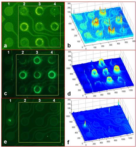 Figure 2 - Results for the DEP separation of single-stranded (ss) and double-stranded (ds) high molecular weight (hmw) DNA from whole blood, and a whole blood (no DNA) control. Discussion and Conclusions: DEP devices and methods have been developed which have allowed the rapid isolation, concentration and detection of both hmw-ss-DNA and hmw-ds-DNA from whole blood samples. Hmw-DNA fragments could be easily detected at clinically relevant levels of 260 ng/ml for ss-hmw-DNA. In other DEP work we were able to show that fluorescent 40nm nanoparticles could be isolated and detected from whole blood at a level of 9.5 x 10⁹ particles/ml. Additionally, using DEP devices we have been able to collect hmw-cfc-DNA from Chronic Lymphocytic Leukemia (CLL) and other cancer patient whole blood samples. Blood sample to PCR could be achieved in less than twenty minutes. Important for future diagnostic applications, these DEP results could be obtained using only very small sample volumes (20 µl whole blood) with no sample preparation being required. Further DEP experiments have demonstrated that very low molecular weight DNA appears not to concentrate into the high-field regions. This could be beneficial in that it shows, at least at the DEP AC voltages and frequencies being used, that the process is more selective for high molecular weight DNA. Thus, DEP may have a selective advantage for the isolation of cancer and other disease-related cfc-DNA, rather than the lower molecular weight apoptotic DNA normally found in blood. In addition to specific fluorescent or colorimetric stains, numerous other molecular biology techniques including PCR, in situ hybridization and immunochemistry detection can be easily carried out in the same sample chamber where isolation of the entities has occurred. Taken together, the results of this study support the enormous potential of DEP as a “seamless sample-to-answer” technique for the rapid isolation and detection of disease related cfc-DNA and nanoparticles directly from whole blood and other complex samples.
See more of this Session: Advances in Electrokinetics and Electrophoresis: Fundamentals
See more of this Group/Topical: Topical 3: 2012 Annual Meeting of the American Electrophoresis Society (AES)
See more of this Group/Topical: Topical 3: 2012 Annual Meeting of the American Electrophoresis Society (AES)