
 Google
 Yelp
 Facebook
 Twitter
 ChEnected
300427 Confinement of a DNA Dodecamer Onto Pristine Carbon Nanotubes: Stability of the Canonical B Form
Confinement of a DNA Dodecamer onto Pristine Carbon Nanotubes and the Stability of the Canonical B Form
Fernando J.A.L. Cruz1,2, Jose P. B. Mota1 and Juan J. de Pablo2,3
1 Requimte/CQFB, Dept. Chemistry, Universidade Nova de Lisboa, Caparica, Portugal. 2 Dept. Chem. Bio. Eng., University of Wisconsin-Madison, Madison, WI, USA. 3 Institute of Molecular Engineering, University of Chicago, Chicago, Illinois, USA.
fj.cruz@fct.unl.pt
<>I. Introduction
Deoxyribonucleic acid (DNA) and single-walled carbon nanotubes (SWCNTs) are prototypical one-dimensional structures; the former plays a central role in chemical biology and the latter holds promise in nanotechnology applications.1-3 From the point of view of biological purposes and DNA manipulation, carbon nanotubes have been proposed as templates for DNA encapsulation, intracellular penetration via endocytosis and delivery of biological payloads and ultrafast nucleotide sequencing. While structure in its natural form and environment is well established (e.g. B-DNA in aqueous solution), their interactions have been the subject of intense investigation, nonetheless, the corresponding molecular-level phenomena remain rather unexplored. Is confinement spontaneous from a thermodynamical point of view (free-energy)? How important are the conformational properties of the double strand upon confinement (entropy)? What is the effect of the media electrical charge density on the negatively charged DNA backbone (ionic strength)? How does the confinement process depend on intrinsic properties of the nanotube such as diameter (D)? Previous theoretical and experimental work has focused almost exclusively on exoadsorption of DNA at the SWCNTs external walls, overlooking the possibility of endohedral confinement; the energetics of the latter is one of the main purposes of the present work. Confinement driven adsorption is a phenomenon that is utterly unmapped, and most of the previous work has been focused in systems at temperatures away from the physiological value and/or ionic strength, thus precluding extrapolation of results to in vivo conditions. We'll address this issue using molecular dynamics and enhanced sampling techniques (metadynamics, umbrella sampling) to probe the encapsulation mechanism of an atomistically detailed DNA dodecamer, onto SWCNTs of different diameters, employing precise physiological conditions, 310 K and [NaCl] = 134 mM.
II. Results and Discussion
Confinement
of DNA onto a (51,0) topology, D =
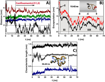
4nm, is fast and becomes complete before 16 ns of observation time (Fig.1A);
initially (t = 0 – 2 ns), the
double strand is in the bulk, and as it diffuses towards the SWCNT undergoes
structural rearrangements leading to minor increases in pitch length, P, and end-to-end distance, L. After 2 ns the dodecamer is already
at the nanopore entrance, experiencing strong van der Waals attractions towards
the solid, leading to complete encapsulation at 15.42 ns, after which it
relaxes and reassumes its unconfined structure (P = 3.4 nm, L = 4.1 nm)
consistent with the geometrical characteristics of B-DNA in bulk.4 It is interesting to observe that
confinement is unalterable, and the biomolecule never returns back to the bulk
solution during the observation time window, always maintaining direct contact
with the solid at a distance of closest approach of ca. 0.26 nm; nonetheless, the confined molecule retains its
translational mobility diffusing along the nanopore main axis (Fig. 1B). In
contrast, the narrowness of a (40,0) topology, D = 3 nm, compared to the DNA skeletal diameter, inhibits
encapsulation even for an observation time of 0.1 μs.
We determined the characteristic lengths, radius of gyration (Rg)
and its projection along the nanotube main axis (


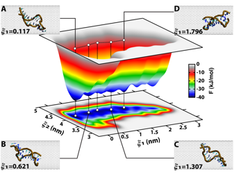
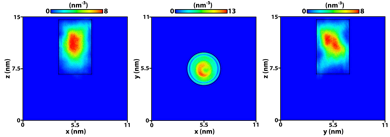
The thermodynamical stability of encapsulation is probed by the free-energy (F) differences associated with the process, using well-tempered metadynamics.5 An inspection of the resulting three-dimensional surface (Fig.2) reveals the existence of five distinct free-energy minima, sharing in common the fact that all are located at discrete positions along the nanopore internal volume, ξ1 < 1.8 nm; the absolute minimum at ξ1 = 0.117 nm evidences the SWCNT center as the energetically most stable region subsequent to encapsulation. In order to escape from those deep free-energy valleys, F ~ – 40 kJ/mol–1, the DNA molecule has to overcome large energetic barriers, rendering the exit process towards bulk solution a thermodynamically expensive one. The number density maps depicted in Fig.3, obtained from atomically detailed mass histograms, corroborate the finding that free-energy is minimum at the nanopore center; Fig.3 also reveals a thin and cylindrically-shaped exclusion volume close to the hydrophobic solid walls, arising from the repulsion between the latter and the heavily charged phosphate groups. It is remarkable to observe that the exact position of the free-energy minima oscillate little around ξ2 = 4.1 nm, coherent with L = 4.1 nm characteristic of a B–DNA structure (Fig.1).
As far as we are aware these observations are the first of their kind, and they come to pave the way for the design of smart nanotube based devices for in vivo DNA encapsulation.
| FIGURE 1 - Encapsulation of DNA onto a (51,0) SWCNT. A) Kinetics. Encapsulation is complete after 15.42 ns and the double-strand never returns back to the bulk solution during the observation time window; DNA maintains its translational mobility within the nanopore. Notice that a 2 nm c.o.m. distance between the biomolecule and the solid corresponds to the threshold bellow which complete encapsulation occurs: black) distance between centres of mass of DNA and (51,0) SWCNT, dark red) distance between centres of mass of DNA and (40,0) SWCNT, green) DNA end-to-end length, blue) DNA pitch length, grey) minimum distance between any DNA atom and the SWCNT. B) State diagram. Distances between terminal nucleobase pairs and the SWCNT entrance, projected along the nanotube main axis: black) terminus 1 and red) terminus 2. The horizontal dashed lines correspond to the SWCNT boundaries and the inset magnification depicts the first instant just after complete encapsulation, representing the terminus 1 atoms in black and the terminus 2 atoms in red. The encapsulation mechanism can be described by a three-step kinetics: I) fast diffusion of bulk DNA towards the nanopore (0 – 2 ns), II) strong van der Waals attraction towards the solid, leading to confinement of terminus 1 at 2.33 ns, and finally III) penetration of terminus 2 onto the nanopore volume resulting in complete encapsulation of DNA. Note the confined molecule maintains its translational mobility along the nanopore axis. C) Characteristic lengths. black) radius of gyration, Rg, grey) radius of gyration z-component,
|

| FIGURE 2 - Free energy landscape of DNA@(51,0)SWCNT hybrid. ξ1 is the distance between centres of mass of the DNA and SWCNT, projected along the nanopore main axis, and ξ2 is the absolute distance between (GC) termini on opposite sides of the double-strand, equivalent to the DNA end-to-end length. The several free-energy minima along ξ1 demonstrate that the molecule is relatively mobile to translocate along the nanotube, however, the absolute minimum at ξ1 = 0.117 nm indicates that the nanopore center is the energetically favoured region. Interestingly, all the ξ1 minima are located along a quasi-linear path defined by ξ2 Å 4.1 nm highlighting the enhanced thermodynamical stability corresponding to the canonical B form. Snapshots were taken at (ξ1, ξ2) nm = A) (0.117, 4.112), B) (0.621, 4.164), C) (1.307, 4.164) and D) (1.796, 4.115).
|
| FIGURE 3 – Density maps of DNA@(51,0)SWCNT. The existence of a cylindrical exclusion volume close to the walls, r Å 0, is the direct consequence of direct repulsion between the heavily charged phosphate groups and the hydrophobic solid. The dashed lines indicate the boundaries of the nanotube.
|
References
(1) H. Kumar et al, Soft Matter 2011, 7, 5898.
(2) B.M. Venkatesan, R. Bashir, Nature Nano. 2011, 6.
(3) A. D. Franklin et al, Nano Lett. 2012, 12, 758-762.
(4) J.M. Vargason et al, Proc. Nat. Acad. Sci. 2001, 98, 7265.
(5) A. Barducci et al, Phys. Rev. Let. 2008, 100, 020603.
See more of this Group/Topical: Nanoscale Science and Engineering Forum