
 Google
 Yelp
 Facebook
 Twitter
 ChEnected
305635 Fabric Microfluidics for Low-Cost Protein Separations
Introduction: Point-of-care diagnostic tests (POCTs) are necessary to screen for disease and monitor treatment at the site of patient care. By this definition, a POC device must be standalone, consume minimum laboratory resources and also be inexpensive [1]. For instance, the typical paper-based POCT for pregnancy consists of a designated path for a small volume of urine to flow along, complex with assay reagents and generate a result in terms of a colored signal that can be interpreted by the lay user. However, the manufacturing process involves multiple coating, drying and alignment steps that can limit scalability and contribute to cost. In prior work, we demonstrated a seamless, woven fabric-based microfluidics platform to address this issue [2]. Weaving allows us to combine reagent patterning and device assembly into a quasi-single step. Two main avenues will be discussed in the context of fabric POCTs: a. Passive control of liquid flow in fabric b. Feasibility of fabric as a platform for the electrophoretic separation of proteins from biological samples.
 Materials and Methods: Fabric samples were weaved on a handloom at
Achira Labs, Bangalore, India. The flow properties that are required to
maneuver medical samples such as serum or urine along a reagent-loaded pathway
are provided by controlled structural in-homogeneities in the fabric. To
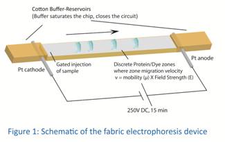
manufacture the electrophoretic chip, Platinum wire electrodes were weaved into
a polyester flow path with absorbent cotton ends as buffer-reservoirs (Figure
1). An Ohm's Law Plot was performed to
ensure that the system could withstand a minimum of 250V DC. In order to
discern diffusion effects from electrophoresis, dyes that can be tracked
visually were run on fabric with and without an applied voltage. After
confirming dye migration in an electric field, a dye-conjugated protein ladder
(ColorBurstTM, Sigma) was resolved in
fabric at 250V and compared to a standard PAGE run.
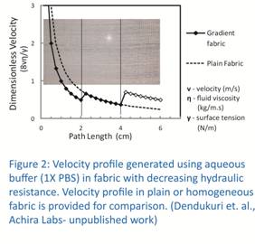 Results and Discussion: Figure 2 shows a saw-toothed velocity
profile generated in a fabric where hydraulic resistance decreases in three
seamless steps. This illustrates that
sample flow can be slowed in regions where time-sensitive reactions must take
place. The colored
protein ladder was resolved by electrophoresis. Future work will attempt to
resolve fluorescent proteins for use with the more accurate and quantitative
detection of fluorescence.
Conclusions: From prior work 1 and from the electrophoretic separation achieved, fabric is a versatile platform for a variety of analytical procedures. Added to the high manufacturing throughput per-day on a standard industrial loom, fabric POCTs can be disseminated at low costs to resource deprived medical laboratories.
References:
1. P. Yager, G. J. Domingo, J. Gerdes (2008). Annu. Rev. Biomed. Eng. 10: 107-144
2. P.Bhandari, T.Narahari, D.Dendukuri (2011). Lab Chip 11(15): 2493-2499.
See more of this Group/Topical: 2013 Annual Meeting of the American Electrophoresis Society (AES)