
 Google
 Yelp
 Facebook
 Twitter
 ChEnected
315724 Using Magnesium Oxide Nanoparticles to Improve Inhomogeneous Soft and Hard Tissue Growth
Introduction: Where ligaments meet bone, there exists a transitional region of inhomogeneous tissue that is graded from hard, highly mineralized fibrocartilage at the bone interface to un-mineralized soft tissue at the ligament. This structure, called the enthesis, disperses stress concentrations that arise due to the vastly different mechanical properties of bone and ligaments. However, in the event that a ligament is injured and requires surgery, the tendon-to-bone insertion site (TBI) must be destroyed and cannot easily be restored due to avascularity in the region and because of consequent slow healing times within the inhomogeneous tissue compared to healing within uniform tissues. It is believed that this loss of enthesis functionality following joint reconstructive surgery is a leading cause of high failure rates for such surgeries (5-25% failure rates for ACL surgery) [1]. Therefore, there is considerable interest in the development of a nanostructured biomaterial that is capable of regenerating the TBI.
The current study developed and characterized for the first time poly (l-lactic acid) (PLLA) mineralized with nanoparticles of hydroxyapatite (HA) and magnesium oxide (MgO) as a tissue scaffold to direct the regeneration of the enthesis. HA constitutes about 60% of the dry mass of bone and has been used previously as a successful material for bone grafts. Magnesium is an essential mineral that regulates HA crystal size and density, and Weng and Webster have shown that nano-rough magnesium increased bone cell density by up to three-fold when compared to bulk magnesium [2]. Presently, the ability of materials to promote tissue growth at the TBI was characterized via cell adhesion and proliferation experiments with fibroblasts and osteoblasts. Further characterization of materials was performed using scanning electron microscopy (SEM), contact angle tests, and mechanical load testing.
Methods: Hydroxyapatite (HA) precipitates were synthesized by a wet chemistry process and then treated hydrothermally to give nanoparticles of sizes consistent with HA crystals observed in natural bone, as reported by Zhang et al. [3]. Poly (l-lactic acid) (PLLA) (MW=50,000), (Polysciences, Warrington, PA) was placed in 20 mL scintillation vials and dissolved in 10 mL of chloroform (Sigma Aldrich, St. Louis, MO) to reach 3 wt% PLLA in chloroform. HA nanoparticles and MgO nanoparticles (particle diameters of 20 nm, US Research Nanomaterials Inc., Houston, TX) were added to separate vials in concentrations ranging from 5-20 wt% in PLLA. Samples were heated to 55 °C and sonicated for 1 hour, then poured into 60 mm diameter pyrex petri dishes (Sigma Aldrich, St. Louis, MO). Samples were heated at 60 °C for 1.5 hours to evaporate excess chloroform, producing polymer films about 0.2 mm thick which were then allowed to sit overnight before being cut into strips for further study.
Samples were cut into 1 cm x 3 cm rectangular strips for tensile testing with a uniaxial tensile tester equipped with material analysis software (ADMET, Norwood, MA). A 10 lb. load cell was used and small pieces of paper towel were attached to the tensile grips to prevent the polymer composites from breaking from the force of the grips. This set-up was used to obtain the elastic modulus, material elongation, and maximum load endured for each sample. Cell adhesion and proliferation tests were performed by seeding 3500 cells/cm2 of fibroblasts and osteoblasts (American Type Culture Collection, Manassas, VA) onto 1-cm2 samples and culturing for 4, 24, 72, and 120 hours under standard conditions. Cell numbers were quantified using MTT assays (Sigma Aldrich, St. Louis, MO). Experiments were conducted in quadruplet and repeated three times. Data was analyzed using Student's t-tests.
Results: Results indicated for the first time that MgO nanoparticles in plain PLLA or PLLA/HA composites significantly increased osteoblast and fibroblast adhesion on PLLA (Figure 1). SEM images showed considerable differences in nanoscale surface topography between samples. However, preliminary contact angle results demonstrated no differences in wettability among all samples. Mechanical tensile testing revealed that the addition of HA nanoparticles to plain PLLA hardened the polymer, reducing the material elongation and increasing its elastic modulus. These results were exaggerated for PLLA mineralized with MgO nanoparticles. The maximum load that the polymer was capable of withstanding was slightly increased for MgO mineralized samples.
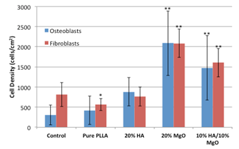
Figure 1. Adhesion of fibroblasts and osteoblasts on pure PLLA and PLLA mineralized with nanoparticles (20% HA, 20% MgO, and 10% HA/10% MgO). Control is empty cell culture well. Data represents mean ± SD. n = 4. % = weight%. *P<0.05, **P<0.005 compared to controls.
Conclusions: Here, MgO nanoparticles have been shown to increase osteoblast and fibroblast cell adhesion on PLLA, and should therefore be further investigated as a material to promote bone tissue growth at one end of the tendon-to-bone insertion site, and fibrous tissue growth at the other end of the TBI.
Acknowledgements: The authors thank Robert Eagan, William Fowle, Scott McNamara, David McKee, the Northeastern University Department of Chemical Engineering for facilities and funding, and the NSF-IGERT grant #0965843 for funding.
References
[1] Smith L, Thomopoulos S, US Muscoskel. Rev., 6 (2011), 11-5.
[2] Weng L, Webster TJ, Nanotechnology. 23 (2012), 485105.
[3] Zhang et al., Int. Journal of Nanomedicine, 3 (2008), 323-34.
See more of this Group/Topical: Nanoscale Science and Engineering Forum