
 Google
 Yelp
 Facebook
 Twitter
 ChEnected
323792 Synchronized Delivery of Vascular Endothelial Growth Factor With Protease Cues Sustains Activation Toward Vessel Formation
Synchronized delivery of vascular endothelial growth
factor with protease cues
sustains activation toward vessel formation
Suwei Zhu, Yunfeng Lu, Tatiana Segura
Chemical & Biomolecular Engineering Department, University of California Los Angeles
Over 6.5 million patients annually in the United States are affected by wound-related health problems resulting from impeded healing processes. Following cutaneous wounding, dermal angiogenesis -- the formation of new blood vessels to bring in nutritive substances and oxygen -- is important for the reconstruction of the normal aspects of skin physiology and yet to be improved in tissue regeneration. More specifically, the signal protein of vascular endothelial growth factor (VEGF), which enhances the survival, proliferation and migration behaviors of endothelial cells, needs to be presented at the wound site in a cell-responsive and sustainable fashion. We have designed a nanocapsule delivery system of VEGF (nVEGF) allowing cargo to be liberated only in the presence of cell-secreted proteases. Unlike conventional protein therapeutic delivery approaches that resort to multi-dose administrations or matrix-assisted retention of growth factors, nVEGF act as a reservoir to sustain modulated release of active VEGF, based on a nanocapsule platform that can be applied alone or within versatile scaffolds.
In our strategy, a nanogel shell comprised of acrylamide based monomers and plamin-sensitive peptide (KNRVK) crosslinker was weaved around VEGF to synthesize protein nanocapsules (nVEGF), which were characterized by transmission electron microscopy (TEM) to be 15~30 nm in diameter, dynamic light scattering (DLS) and enzyme-linked immunosorbent assay (ELISA) for targeted release (Fig. 1). In the aspect of molecular biology, nVEGF altered the phosphorylation profile of VEGF receptor-2 on human umbilical endothelial cells (HUVECs) and downstream signalings into a prolonged activation state significantly different than naked VEGF did (Fig. 2). A three-dimensional in vitro assay of HUVEC sprouting and lumen formation showed that one dose of plasmin-degradable nVEGF supported such behaviors of HUVECs while naked VEGF was soon depleted and supplemental additions were required (Fig. 3). Ex vivo application of nVEGF on the chicken chorioallantoic membrane stimulated the formation of capillaries (Fig. 4). In mouse skin wound healing model, the application of nVEGF within fibrin matrices promoted a faster wound closure with a significantly higher density of infiltrated endothelial cells and capillaries (Fig. 5).
Our results demonstrated plasmin-degradable VEGF nanocapsules sustained the activity of VEGF (preventing clearage and degradation) and achieved the same degree of angiogenesis as re-dosing with exogenous VEGF. Here enzymatically-triggered release of VEGF from the nanocapsule reservoir synchronized the dosage with tissue endogenous protease levels. By reducing the frequency of therapeutic administration, such applications of growth factor nanocapsules would be clinically relevant and practically feasible to continuous deliver active growth factors in regions with proteases and at times of demand.
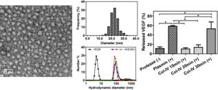
Figure 1 Characterization of the size of nVEGF and the degradability of its nanogel shell to release cargo VEGF. TEM image of purified nVEGF showing spherical particle morphology (left) and measured distribution of nanocapsule diameters (N = 322) (upper center). Hydrodynamic sizes of different batches of nVEGFs and naked VEGF (lower center). Release of VEGF from nVEGF upon protease degradation of nanogel shells (n = 3; mean ± SEM) (right).
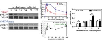
Figure 2 nVEGF prolongs the phosphorylation of VEGFR-2 (Tyr 1175) due to VEGF release by cell-produced proteases. Western blot analysis from HUVECs exposed to native VEGF and nVEGF for different lengths of time (left) and normalized phosphorylation profiles (n = 3; mean ± SEM) (upper center). Phospho-ELISA analysis of HUVECs exposed for 30 min to consecutively reapplied VEGF and nVEGF (right) and the prolonged phosphorylation profile reproduced by additions of a small amount of VEGF in the last 5 min of each incubation period (lower center) (n = 3; mean ± SEM).
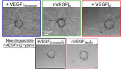
Figure 3 Single dose plasmin-degradable nVEGF supports the sprouting of HUVECs in fibrin matrices over 7 days in the bead assay.
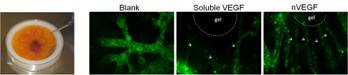
Figure 4 Ex vivo implantation of fibrin matrices with VEGF or nVEGF induces capillaries in chicken chorioallantoic membrane assay.
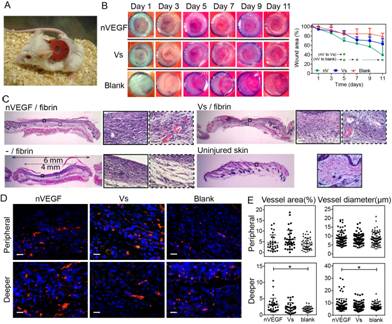
Figure 5 Mouse wound healing model and angiogenesis.
See more of this Group/Topical: Food, Pharmaceutical & Bioengineering Division