
 Google
 Yelp
 Facebook
 Twitter
 ChEnected
325844 Fabrication of Jell-O Milli-Fluidic Chips for Hands-On Education of Hemodynamics and Blood Cell Adhesion
Fabrication of Jell-O Milli-fluidic Chips for Hands-On Education of Hemodynamics and Blood Cell Adhesion
Michael J. Mitchell1, David Syracuse2, Carlos A. Castellanos1, Shivaun D. Archer1, Michael R. King1
1Department of Biomedical Engineering, Cornell University, Ithaca, New York USA
2TST BOCES High School, Ithaca, New York USA
Introduction: Advances in microfluidics have allowed for the development of devices that can recreate the complex microenvironment of the bloodstream. In particular, our lab has recently developed microscale flow devices that mimic postcapillary venules to study the basic mechanisms of cancer metastasis (1) and inflammation (2,3). Such devices have also been adapted to kill cancer cells in the bloodstream (4), and isolate circulating tumor cells from patient blood (5). As the clinical benefits of microfluidics are becoming apparent, it is essential to educate young students on basic fluidic principles and their potential impact on human disease diagnosis and treatment. A simple fabrication technique was demonstrated using Jell-O to teach high school students the essential components of blood flow, and its subsequent effects on cell adhesion in inflammation and cancer.
Materials and Methods: Model milli-fluidic devices were fabricated using a soft lithography technique consisting of foam plates, coffee stirrers, single- and double-sided tape, and Jell-O. Briefly, coffee stirrers were cut, connected, and arranged in various formations and adhered to foam plates using double-sided tape. Single-sided tape was placed on top of the coffee stirrers to create a smooth surface. A Jell-O liquid was prepared as per manufacturer's instructions, poured onto molds, and left to cure inside a refrigerator for 2 days. Jell-O chips were then peeled from molds and placed onto aluminum plates. Inlets and outlets were created within the chips, and liquid was perfused through the device using disposable transfer pipets. For blood cell adhesion experiments, chips were peeled and placed onto glass slides, and incubated with a solution of soluble E-selectin protein for 2 hours. Human neutrophils were perfused through E-selectin coated Jell-O chips, and assessed for cell adhesion using brightfield microscopy.
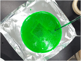
Results and Discussion: High school students successfully fabricated model milli-fluidic devices consisting of varying fluidic networks (Figure 1). Comparisons of Jell-O chip versus polydimethylsiloxane (PDMS) microfluidic chip fabrication in biomedical research were highlighted throughout the fabrication process. The concept of pressure-driven flow was demonstrated using Jell-O chips in the absence of an outlet, as fluid could not flow due to air present in the channel, which could not escape due to the absence of an outlet. High school students calculated flow rate by measuring the flow of a set volume of fluid through the device over time (Figure 2). The concept of laminar flow was demonstrated to students via perfusion of both a light and a dark-colored fluid through a Y-shaped Jell-O chip, as fluids perfused through channels simultaneously did not mix. Jell-O chips were also used to demonstrate cell adhesion, as E-selectin coated devices successfully induced human neutrophil adhesion. Graduate students, so to prevent contamination and exposure of hazardous materials to high school students, demonstrated all cell adhesion studies.
Conclusions: We have shown that simple, inexpensive Jell-O microfluidic devices can successfully demonstrate the basic principles of blood flow in a hands-on high school laboratory setting. The fabrication and use of devices can benefit students by introducing them to current biomedical technologies and applications, and encourage the pursuit of engineering and science degrees at the university level.
Acknowledgements: This study was supported by a National Science Foundation GK-12 grant DGE:0841291.
Figure 1: Schematic to fabricate Jell-O chips using soft lithography. (A) Top view of a negative mold consisting of coffee stirrers and a foam plate. (B) Side view of coffee stirrers adhered to foam plate using tape. (C) Liquid Jell-O is poured onto the mold. (D) Chip is solidified over using refrigeration and then peeled from plate. (E) Chip is adhered to aluminum plate or foil for use.
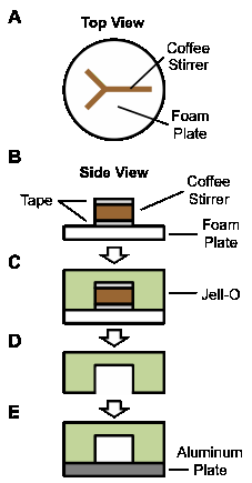
Figure 2: Perfusion of fluid through a Jell-O chip.
References:
1. Yin X, Rana K, Ponmudi V, King MR. Knockdown of fucosyltransferase III disrupts the adhesion of circulating cancer cells to E-selectin without affecting hematopoietic cell adhesion. Carbohydrate Research. 2010;345:2334–42.
2. Mitchell MJ, King MR. Shear-Induced Resistance to Neutrophil Activation via the Formyl Peptide Receptor. Biophysical Journal. 2012 Apr 18;102:1804–14.
3. Cao TM, Takatani T, King MR. Effect of extracellular pH on selectin adhesion: theory and experiment. Biophysical Journal. 2013 Jan 22;104(2):292–9.
4. Mitchell MJ, Chen CS, Ponmudi V, Hughes AD, King MR. E-selectin liposomal and nanotube-targeted delivery of doxorubicin to circulating tumor cells. Journal of Controlled Release. 2012 Jun;160:609–17.
5. Hughes AD, Mattison J, Western LT, Powderly JD, Greene BT, King MR. Microtube Device for Selectin-Mediated Capture of Viable Circulating Tumor Cells from Blood. Clinical Chemistry. 2012 Feb 16;58:846–53.
See more of this Group/Topical: Education Division