
 Google
 Yelp
 Facebook
 Twitter
 ChEnected
326240 Unnatural Killer Cells: TRAIL-Coated Leukocytes That Kill Cancer Cells in the Circulation
Unnatural Killer Cells: TRAIL-coated Leukocytes that Kill Cancer Cells in the Circulation
Michael J. Mitchell1, Kuldeepsinh Rana1, Elizabeth C. Wayne1, Chris B. Schaffer1, Michael R. King1
1Department of Biomedical Engineering, Cornell University, Ithaca, New York USA
Introduction: Hematogenous metastasis contributes to a poor patient prognosis in many types of cancer, with over 90% of cancer-related death attributed to metastasis (1). For metastasis to occur, cancer cells detach from the primary tumor, invade through the basement membrane, and intravasate into the peripheral circulation as circulating tumor cells (CTCs) (2). It is believed that adhesive interactions between selectins on the blood vessel wall and selectin ligands on the CTC surface facilitate metastatic progression, in a manner similar to the leukocyte adhesion cascade essential in the inflammatory response (3). Here, we describe a novel approach to functionalize human and murine leukocytes with tumor necrosis factor-related apoptosis-inducing ligand (TRAIL) on their surface, along with E-selectin (ES), to capture and induce cancer cell apoptosis both in vitro in human blood and in vivo in mouse circulation.
Materials and Methods: ES/TRAIL nanoscale liposomes and COLO 205 or PC-3 cancer cells suspended in buffer or spiked in peripheral human blood were subjected to shear flow in a cone-and-plate viscometer. Cancer cells and mononuclear cells were isolated, cultured overnight, and analyzed for viability using flow cytometry (FC). To determine leukocyte functionalization with ES/TRAIL, fluorescent (FL) liposomes were exposed to human blood under shear flow, and leukocytes were isolated and analyzed for ES/TRAIL attachment using FC and confocal microscopy. To examine cancer cell capture and killing in vivo, mice were injected with ES/TRAIL retro-orbitally, followed by injection of FL-labeled COLO 205 cells via tail vein injection. Mouse blood was removed via cardiac puncture, and viable and apoptotic cancer cells were analyzed using FC. Viable and apoptotic cancer cells lodged within the mouse lung, liver, and lymph nodes were quantified using multiphoton microscopy.
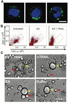
Results and Discussion: Upon exposure to shear flow, ES/TRAIL liposomes in human blood were functionalized to the surface of leukocytes, as confirmed by confocal microscopy (Fig. 1A) and FC. Exposure of functionalized ES/TRAIL leukocytes into cancer cell-spiked blood successfully targeted and killed cancer cells under shear flow, compared to controls (Fig. 1B). Mouse studies revealed that ES/TRAIL-functionalized leukocytes capture and induce apoptosis in cancer cells in the peripheral circulation in vivo (Fig. 1C). Cancer cells lodged within the mouse lung were apoptotic after treatment with ES/TRAIL leukocytes, as determined by multiphoton microscopy.
Conclusions: We have shown that leukocytes functionalized with ES/TRAIL can capture and induce cancer cell apoptosis in vitro in human blood and in vivo in mouse experiments. ES/TRAIL-functionalized leukocytes can form the basis of a novel method to target CTCs in the bloodstream as a means to prevent cancer metastasis. Clinically, this therapeutic strategy can serve as preventive measure upon diagnosis of metastatic hematogenous cancers.
Acknowledgements: The work described was supported by the Cornell Center on the Microenvironment and Metastasis through Award Number U54CA143876 from the National Cancer Institute.
Figure 1: (A) Functionalized ES/TRAIL leukocytes after ES/TRAIL liposome exposure to human blood under shear flow. Green = FL ES/TRAIL liposomes. Blue = leukocyte nuclei. Scale bar = 5 um. (B) Viable COLO 205 cancer cells (gated FC population) after exposure leukocytes functionalized with ES or ES/TRAIL. Untreated: viable COLO 205 control sample. (C) COLO 205 cells (yellow arrow) and functionalized leukocytes (red arrow) isolated from mouse blood after exposure to ES/TRAIL or ES liposomes, sTRAIL, or buffer injections in vivo. Scale bar = 20 um.
References:
1. Mitchell MJ, King MR. Computational and experimental models of cancer cell response to fluid shear stress. Frontiers in Oncology. 2013 Mar 2;:1–11.
2. Hughes AD, King MR. Nanobiotechnology for the capture and manipulation of circulating tumor cells. WIREs Nanomed Nanobiotechnol. 2011. Dec 7;4(3):291–309.
3. Geng Y, Marshall JR, King MR. Glycomechanics of the Metastatic Cascade: Tumor Cell–Endothelial Cell Interactions in the Circulation. Ann Biomed Eng. 2011 Nov 19;40(4):790–805.
See more of this Group/Topical: Food, Pharmaceutical & Bioengineering Division