
 Google
 Yelp
 Facebook
 Twitter
 ChEnected
326340 Submillisecond Pulses of Fluid Shear Stress Suppress Chemoattractant-Induced Neutrophil Activation
Submillisecond Pulses of Fluid Shear Stress Suppress Chemoattractant-Induced Neutrophil Activation
Michael J. Mitchell1, Michael R. King1
1Department of Biomedical Engineering, Cornell University, Ithaca, New York USA
Introduction: Fluid shear stress (FSS) is a ubiquitous component of the microenvironment for leukocytes, as they carry out the physiological processes of initial adhesion to the endothelium, the formation of pseudopods, and migration into tissues (1). Our lab has recently shown that leukocytes exposed to low venular fluid shear stresses (0.1-4.0 dyn/cm2) develop a resistance to chemoattractant-induced activation (2). However, leukocytes can transiently encounter FSS values >1000 dyn/cm2 in the heart, near the walls of large blood vessels, and at vessel bifurcations. Herein, we evaluated the effect of submillisecond FSS pulses on the neutrophil response to chemoattractants.
Materials and Methods: Neutrophils isolated from peripheral human blood following informed consent were exposed to 1-10 submillsecond FSS pulses via perfusion through microscale conduits using a high-pressure syringe pump, followed by exposure to the chemoattractant formyl-methionyl-leucyl-phenylalanine (fMLP). Expression and spatial distribution of L-selectin, activated αMβ2 integrins, and formyl peptide receptors (FPR) were analyzed using flow cytometry and confocal microscopy. Neutrophil morphology and the appearance of pseudopods were imaged using brightfield and phase contrast microscopy, with images used to measure neutrophil shape factor using an edge detection function. Expression of phosphorylated proteins including p38 MAPK and c-Abl were determined using a Bio-Plex phosphorylated protein detection system.
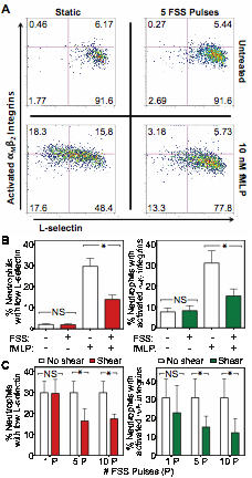
Results and Discussion: In the absence of fMLP stimulation (Fig. 1A), neutrophils exhibited minimal activation when exposed to either static conditions or 5 submillisecond FSS pulses (wall shear stress (WSS) = 6400 dynes/cm2, exposure time = 0.89 ms). Neutrophil samples exposed to static conditions followed by 10 nM fMLP stimulation resulted in a 36% loss of L-selectin and 34% αMβ2 integrin activation (Fig. 1A). However, neutrophil exposure to 5 submillisecond FSS pulses followed by stimulation with 10 nM fMLP reduced L-selectin loss to 16%, and αMβ2 integrin activation to 9% (Fig. 1A). No significant difference in L-selectin shedding and αMβ2 integrin activation was found between sheared and nonsheared neutrophils, whereas a significant reduction in fMLP-induced L-selectin shedding and αMβ2 integrin activation was found in neutrophils exposed to FSS pulses (Fig. 1B). Neutrophil resistance to fMLP-induced activation was FSS pulse-dependent, as 5 pulses of FSS were required to induce neutrophil resistance to activation (Fig. 1C).
Conclusions: We have shown that brief exposure to submillisecond pulses of FSS can induce a mechanosensitive response in human neutrophils. Exposure to high FSS pulses reduce neutrophil activation in the presence of bacterial peptides, and likely serve as a physiological regulator of neutrophil activation and invasion into tissues. Insight into the molecular basis of the neutrophil response can reveal important drug targets for inflammation and hemostasis.
Acknowledgements: This study was supported by the National Institutes of Health, grant No. HL018208.
Figure 1: Submillisecond FSS pulses suppress fMLP-induced neutrophil activation. (A) Neutrophils exposed to static conditions or 5 submillisecond pulses of FSS (WSS= 6400 dyn/cm2, pulse time = 0.89 ms) followed by 10 nM fMLP treatment. As indicators of activation, neutrophils were labeled for L-selectin expression and αMβ2 activation and analyzed using flow cytometry. (B) Neutrophil L-selectin shedding and αMβ2 activation from n = 4 donors, when exposed to FSS and/or fMLP. (C) Neutrophil fMLP-induced L-selectin shedding and αMβ2 activation after exposure to 1-10 submillisecond FSS pulses.
References:
1. Springer TA. Traffic signals for lymphocyte recirculation and leukocyte emigration: The multistep paradigm. Cell. 1994 Jan;76(2):301–14.
2. Mitchell MJ, King MR. Shear-Induced Resistance to Neutrophil Activation via the Formyl Peptide Receptor. Biophysical Journal. 2012 Apr 18;102:1804–14.
See more of this Group/Topical: Food, Pharmaceutical & Bioengineering Division