
 Google
 Yelp
 Facebook
 Twitter
 ChEnected
330503 DEP Isolation of Cancer Related Circulating Cell Free (CCF) DNA Biomarkers Directly From Blood
AIChE 2013
Topical 3: 2013Annual Meeting of the American Electrophoresis Society
“T3006 – “Electrokinetics for Sample preparation”
DEP Isolation of Cancer Related Circulating Cell Free (CCF) DNA Biomarkers Directly from Blood
Michael J. Heller1,2,*Avery Sonnenberg1, Jennifer Y. Marciniak1, Laura Rassenti3, Emanuela Ghia3, Elaine Skowronski1, Sareh Manouchehri1, George Widhopf3 and Thomas J. Kipps3
University of California San Diego, Department of Bioengineering1, Department of Nanoengineering2, and UCSD Moores Cancer Center3, La Jolla, CA, USA 92093-0448
Abstract – Circulating cell free (ccf) DNA/RNA biomarkers are now widely used for cancer diagnostics and management. Unfortunately, the isolation of ccf-DNA/RNA directly from blood has not been possible, and its isolation from plasma is a long and involved process. We report on an AC electrokinetic approach for the rapid isolation of ccf-DNA directly from a small volume of un-processed blood. A dielectrophoretic (DEP) microarray device (chip) was used to isolate ccf-DNA directly from fresh blood samples of fifteen Chronic Lymphocytic Leukemia (CLL) patients and three healthy individuals. Ccf-DNA/RNA from 25uls (a drop) of blood was separated and concentrated into DEP high-field regions in about three minutes, while blood cells, proteins and other biomolecules were removed by a fluidic wash. Concentrated ccf-DNA (SYBR-Green stained) was detected by fluorescence and then eluted for PCR and DNA sequencing. The complete process, blood sample to ccf-DNA for PCR, was completed in less than 10 minutes. Eluted ccf-DNA, from 5ul of the original CLL blood sample, was amplified by PCR using IGHV-specific primers to identify the unique IGHV gene expressed by the leukemic B-cell clone (ie the major IGHV subgroups expressed by CLL B cells IGHV1, IGHV3, IGHV4). The PCR results obtained by DEP from CLL blood were comparable to results obtained using conventional sample preparation of ccf-DNA which starts with one ml of plasma. The DNA sequencing results from the PCR amplified cff-DNA obtained by DEP correlated with earlier sequencing results from genomic DNA isolated from the CLL patient's leukemic B cells. The ability of DEP to provide rapid isolation and detection of ccf-DNA from the equivalent of a drop of blood represents a major step forward in the quest for “liquid biopsy” and point of care (POC) cancer diagnostics and patient monitoring.
Introduction - AC electrokinetic methods like dielectrophoresis (DEP) are well-known techniques for achieving separations of cells, nanoparticles, and biomolecules. However, until recently DEP technology had remained impractical for general use with biological/clinical samples (blood, plasma, serum) and in high conductance buffer solutions (>1mS/cm). For example, earlier DEP work on separating bacteria from blood (~7-9 mS/cm) required a 50-fold dilution of the blood sample before the DEP separation process could be carried out [1]. More recently, we have developed new DEP devices and methods that allow hmw-DNA, nanoparticles, bacteriophage and circulating cell free (ccf) DNA to be isolated and detected from both high ionic strength buffers and blood [2-6]. Using special microarrays with robust platinum microelectrodes over-coated with novel hydrogel layers (designed and fabricated by Biological Dynamics, La Jolla, CA) we are now able to demonstrate the rapid isolation, fluorescent detection and subsequent PCR amplification and DNA sequencing of cancer related ccf-DNA directly from a 25 ul sample of un-processed blood from Chronic Lymphocytic Leukemia (CLL) patients.
Results: Figure 1 (below) shows the electrokinetic DEP device (Biological Dynamics) used for the separation of ccf-DNA from CLL patient blood. The DEP microarray device/chip is a 10 x 12 mm silicon die patterned structure with an interdigitated array of 1000 (60 µm dia.) platinum microelectrodes on 160 um center-center pitch. The microarray is over-coated with a 200nm-500nm porous hydrogel layer. A printed circuit board is attached to the chip using electrically conductive pressure sensitive adhesive which provides electrical contact to standard pogo pin connectors. The PCB cutout forms the sides of the flow cell and an acrylic window with fluidic input and output ports forms the top cover, resulting in a 20 ul flow cell volume. Typically, ~25 ul of the sample would be aspirated to completely fill the flow cell and the fluidic ports. For the actual process, the chip was first pre-treated with approximately 25 ul of 0.5X PBS and run at 2Vrms, 5Hz sine waveform for 15 second to improve the hydrogel porosity. The fluid was aspirated and 25 ul of undiluted sample (blood) was then applied to the chip flow cell and run at 12 Volts peak to peak (Vp-p) at 10 kHz (sine wave) for 3 minutes.
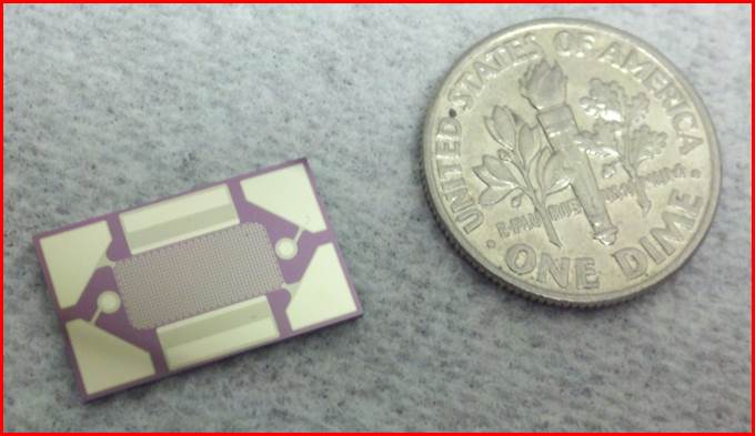 Figure 1 – Electrokinetic DEP
microarray chip device designed and fabricated by Biological Dynamics for
carrying out separations in high conductance buffers and clinical samples
(blood, plasma, serum, etc.)
A checker board field geometry is formed by the AC bias with the positive DEP high-field regions occurring on the microelectrodes, and negative DEP low-field regions occurring between microelectrodes. With the AC voltage ON, the flow cell was washed with TE buffer for 5 minutes at 100 ul/min flow rate. After the wash step, the power was turned OFF allowing the captured DNA to diffuse off the hydrogel. The sample was then aspirated from the flow cell and stored at 4o C in a microcentrifuge tube. In order to visualize real time capture on the microelectrode array, the CLL and normal blood samples were treated with 5x SYBR Green I fluorescent dye which stains the double stranded DNA. Bright field and fluorescent images of the microelectrode pads were then acquired using a CCD camera with a 10x objective, FITC fluorescent filters, and a 470nm LED excitation source. Figure 2 shows representative fluorescent images of the SYBR Green stained ccf-DNA from one of the normal blood samples along with two CLL patient blood samples. MATLAB was used to create 3D fluorescent intensity images which provide better quantification of the relative amount of ccf-DNA that has been concentrated onto the microelectrodes by the DEP process. In general, fluorescent DNA levels were higher in all 15 CLL patient samples than in the normal blood samples; and in most cases they were significantly higher. The concentration of ccf-DNA in the eluted samples (25ul) was also determined by PicoGreen fluorescent analysis. Again the fluorescent DNA levels were higher in all 15 CLL patient samples than in the normal blood samples, and in most cases the concentrations were significantly higher. Subsequent PCR amplification for ccf-DNA (equivalent to 5ul blood) isolated by DEP from all fifteen CLL blood samples compared well with the results obtained from ccf-DNA isolated by the Qiagen process starting with 1ml of CLL patient plasma. Finally, DNA sequencing carried out on the PCR amplified ccf-DNA isolated by DEP correlated very well for all 15 CLL patient samples with sequencing results (Gold Standard) from genomic DNA obtained from the patients B-lymphocyte cells which was isolated from 5 ml of blood.
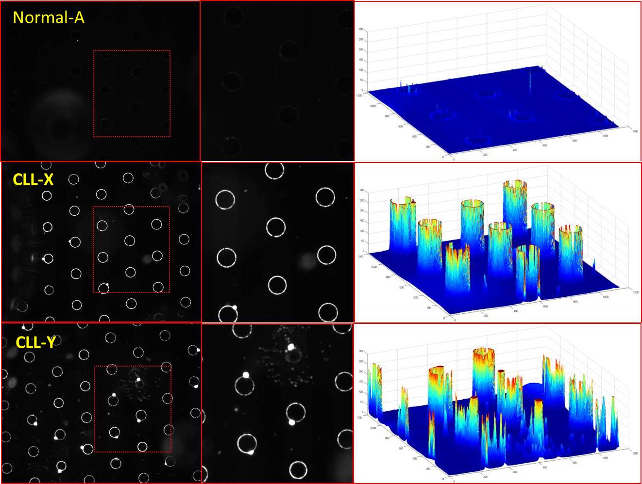 Figure 2 - Fluorescent images of
the SYBR green stained ccf-DNA from a normal blood sample and two CLL patient
blood samples. Also shown (far left) are 3D fluorescent intensity images
created by MATLAB.
Discussion and Conclusions
The use of circulating cell-free (ccf) DNA biomarkers for “liquid biopsy” will unquestionably be important for future point of care (POC) cancer diagnostics and management. Unfortunately, the time and process complexity required for the isolation of ccf-DNA from plasma by conventional methods is a major limitation for developing POC diagnostic formats. Additionally, these processes add significant cost to the assays which will also limit their use. We have now shown that an AC electrokinetic DEP microarray device can be used to isolate ccf-DNA directly from CLL patient blood in less than 10 minutes. The DEP process is not only significantly faster than the conventional methods, but requires only three steps. We have also shown that DEP allows ccf-DNA to be isolated from a relatively small sample volume of only 25 uls, which is equivalent to a drop of blood.
References
(1) Cheng J, Sheldon EL, Wu L, Uribe A, Gerrue LO, Carrino J, Heller MJ and O'Connell JP, “Preparation and Hybridization Analysis of DNA/RNA from E. coli on Microfabricated Bioelectronic Chips”, Nature Biotechnology, Vol. 16, pp. 541-546, 1998.
(2) Krishnan R., Sullivan B.D., Mifflin R.L., Esener S.C., & Heller M.J., AC electrokinetic separation & detection of DNA nanoparticles in high-conductance solutions, Electrophoresis 29, 1765-1774, 2008
(3) Krishnan R. & Heller M.J., AC electrokinetic method for enhanced detection of DNA nanoparticles, J. Biophotonics 2, 253-261, 2009
(4) Krishnan R., Dehlinger D.A., Gemmen G.J., Mifflin R.L., Esener S.C., & Heller M.J., Interaction of nanoparticles at the DEP microelectrode interface, Electrochem. Comm. 11, 1661-1666, 2009
(5) Sonnenberg, A, Marciniak, JY, Krishnan, R, Heller MJ, “Dielectrophoretic Isolation of DNA and Nanoparticles form Blood”, Electrophoresis V33, 2482-2490, 2012
(6) Sonnenberg, A, Marciniak JY, McCanna J, Krishnan R, Rassenti L, Kipps TJ and Heller MJ “Dielectrophoretic Isolation and Detection of cfc-DNA Nanoparticulate Biomarkers and Virus form Blood”, Electrophoresis, V34, pp.1076-1084, 2013
See more of this Group/Topical: 2013 Annual Meeting of the American Electrophoresis Society (AES)