
 Google
 Yelp
 Facebook
 Twitter
 ChEnected
331480 The Improved Adhesion Behavior of Fibroblasts On Anodized 316L Stainless Steel
aCollege of Chemistry, Chemical Engineering and Biotechnology, Donghua University, Shanghai, China
bDepartment of Chemical Engineering, College of Engineering, Northeastern Universtiy, Boston, MA, USA
E-mail: th.webster@neu.edu, Tel: +1 617 373 8565;
Introduction: It is now recognized that surface nanostructures on biomaterials greatly influence cellular behaviors as well as surface biochemical properties [1, 2]. Nano-featured surfaces might be used as an excellent bioactive interface for implantable materials. Stainless steel is one of the most important alloys for biomedical applications. However, their bioinert characteristic surface is a disadvantage for numerous applications. The aim of this study was to fabricate nano-structured surfaces on 316L stainless steel by an anodization method and examine the effects of these anodized nano-surfaces on the attachment of fibroblasts.
Materials & Methods: 316L stainless steel (2.0 mm °Á 2.0 cm °Á 0.5 mm) (Goodfellow Cambridge Ltd. England) was anodized at 30 V for 4 min in ethylene glycol containing 10 vol. % perchloric acid to create bioactive surfaces. The surface morphology and elemental composition of 316 L after anodization were characterized by scanning electron microscopy (SEM, Hitachi S-4800, Tokyo, Japan) and energy-dispersive X-ray analysis (EDAX). Fibroblasts were seeded on the samples to evaluate initial cellular responses to the nano-structured surfaces. After cell culturing for 4 hours, the attachment of fibroblasts was determined by a methyl thiazolyl tetrazolium (MTT) assay. Smoothly polished 316L was used as control surfaces. Light microscopy and SEM were used to observe the morphology of cells. Experiments were completed in triplicate and repeated at least three times. Data were analyzed statistically using student t-test.
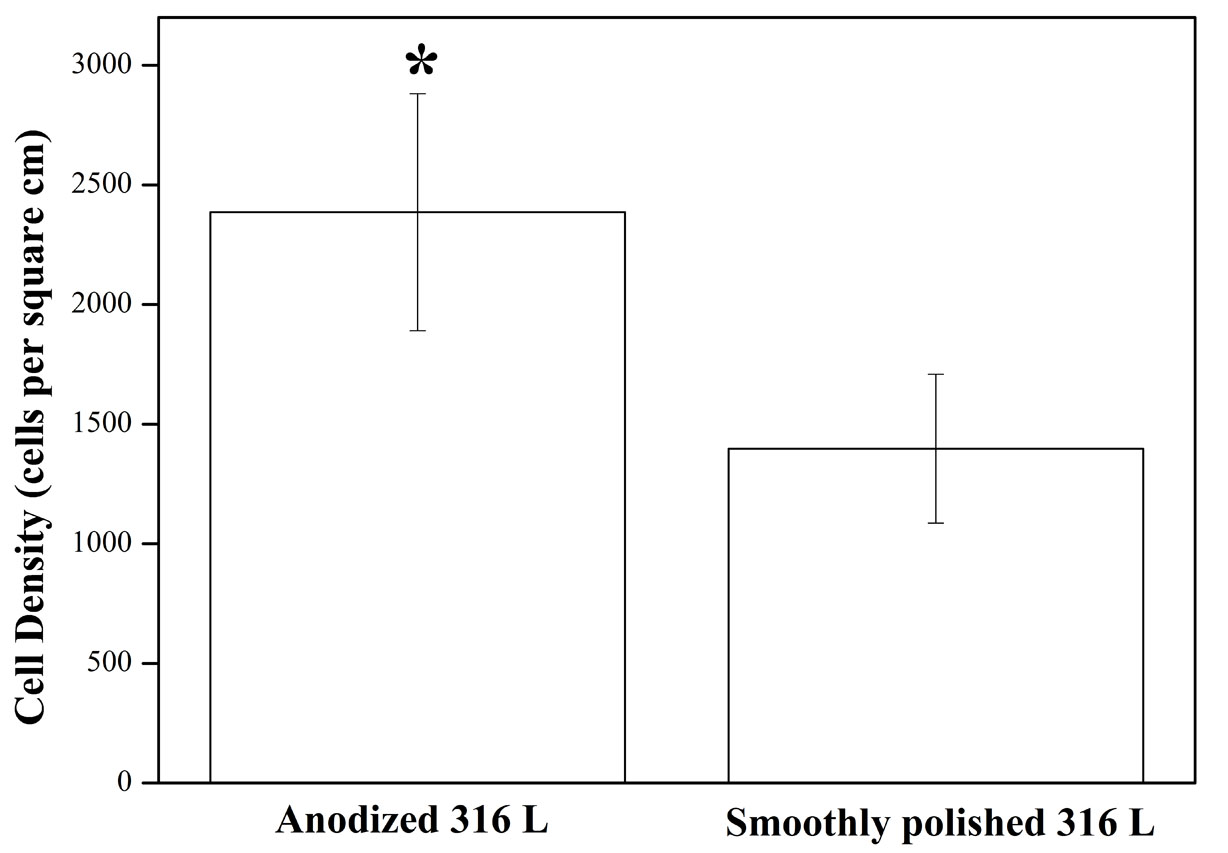
Results and Discussion: A nano-porous structure was successfully fabricated by the anodization procedure. The pore sizes were about 100 nm. After 4 hours of culture, the fibroblasts adhered well on the surfaces of all the stainless steel specimens. SEM images showed that fibroblast exhibited normal morphology and were able to penetrate into pores by long filopodia. MTT tests indicated that the anodization of 316 L stainless steel significantly promoted a higher degree of cell adhesion as compared to the polished 316L stainless steel after 4 hours. (p<0.05) (Fig. 1).
Conclusions: The findings in this study showed that anodization significantly improved the initial cell-material interactions on the 316L stainless steel. Although further work is required to determine the long-term effects of the anodized surfaces on the proliferation and differentiation of cells, the results of this study indicated that the anodized surfaces of the 316L stainless steel had better biological activity.
| Fig.1 The fibroblast attachment on the anodized surface of 316 L stainless steel using the MTT assay for 4 hours. *p<0.05 compared to the smoothly polished 316 L at the same time period |
References
1. Mart¨ªnez E, Engel E, Planell, JA, Samitier, J. Ann Anat 2009; 191: 126-135.
2. Ercan B, Taylor E, Alpaslan E, Webster TJ. Nanotechnology 2011; 22: 295102
See more of this Group/Topical: Nanoscale Science and Engineering Forum