
 Google
 Yelp
 Facebook
 Twitter
 ChEnected
332830 High Aspect Ratio Hydrogel Microwell Arrays As a Novel Platform for the In Situ Cloning of Rare Cells
Introduction: The colony forming unit assay (CFU) for adherent cells is a well established technique for assessing the prevalence and function of rare cells in complex cell mixtures. However, cell migration and colony merger during expansion means the clonality of individual colonies cannot be assured, and the effect of overall plating density on the function, survival and proliferation of rare cells cannot be assessed over a relatively low plating density. We address these shortcomings in the CFU assay by designing high aspect ratio hydrogel microwell arrays which provides an optically transparent and cytocompatible cell growth surface for the attachment and proliferation of cells which are separated by non-fouling PEG walls. This platform enables the in situ analysis of single cell behavior during culture.
Materials and Methods: Non-fouling hydrogel microwell arrays were fabricated through the UV-initiated free radical polymerization of poly(ethylene glycol) dimethacrylate (Mn = 1000) using soft lithography. The arrays have been fabricated on a variety of substrates: glass via silane chemistry, plastics (Mylar and polystyrene) via an argon plasma treatment, and layer-by-layer polyelectrolyte films. Arrays were seeded with bone marrow-derived and expanded aMSCs. Time-lapse video microscopy was used to study single cell migration and proliferation.
Results and Discussion: Hydrogel microwell arrays are stable for greater than two weeks in culture, and the dimensions of the microwells can be easily adjusted by changing the geometry of the PDMS stamp resulting in a versatile cell culture system. Interestingly, aMSCs were able to adhere to and migrate on otherwise cell resistant hydrogels which contact PDMS during molding illustrating a previously unreported phenomena by which micromolding with PDMS undermines the cell resistance of non-fouling hydrogels. We present techniques for overcoming this unwanted cellular attachment. Microwell arrays were fabricated on a range of cell culture substrates (silanized glass, plastics, and polyelectrolyte thin films) and we determined that the substrate affected the fidelity of pattern transfer and the performance of the microwell arrays in containing the seeded cells. Time lapse video microscopy was used to verify that regions within the hydrogel array are able to confine cells and their progeny, and these videos were used to quantify the individual cell proliferation kinetics of aMSCs.
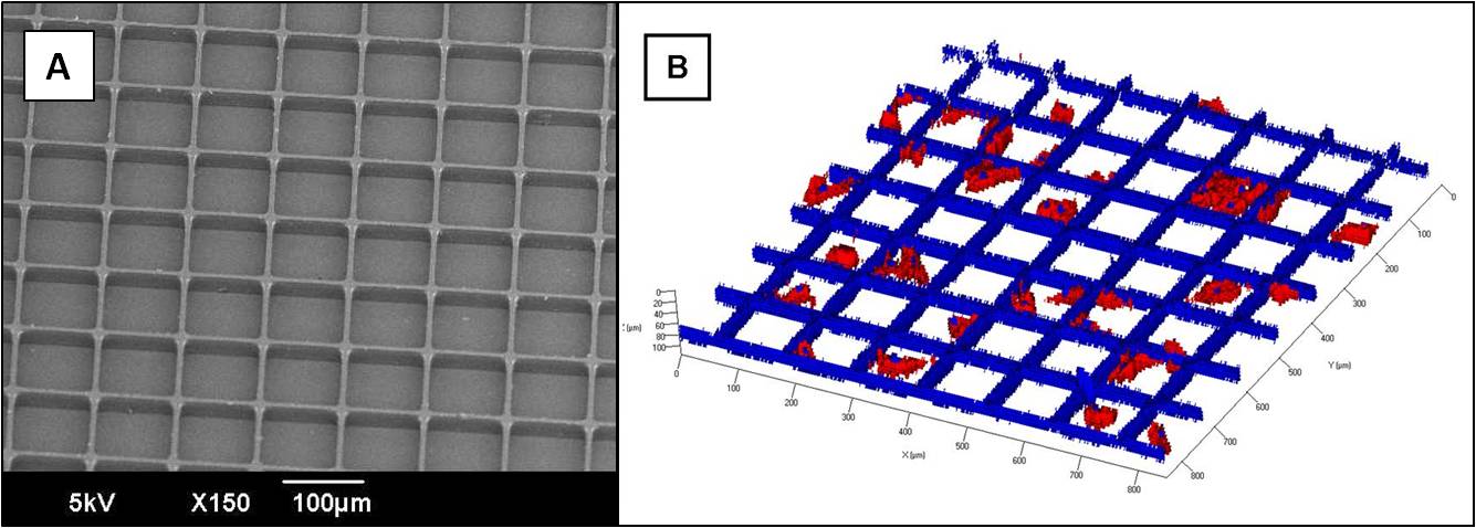
Figure 1. High aspect ratio hydrogel microwell arrays: (A) Scanning electron micrograph showing high fidelity patterning of microwell arrays to polystyrene and (B) confocal microscopy image of mesenchymal stem cells cultured in the hydrogel microwell arrays showing varying proliferation capacities.
Conclusions: High aspect ratio hydrogel microwell arrays which are stable for up to 2 weeks in culture conditions can be fabricated via soft lithography. PDMS stamping alters the non-fouling interfacial properties of PEG, and the cell culture surface affects the quality of pattern transfer during micromolding. Regions of the microwell arrays are capable of containing their cells and their progeny enabling the high resolution study of individual cells behavior.
See more of this Group/Topical: Materials Engineering and Sciences Division