
 Google
 Yelp
 Facebook
 Twitter
 ChEnected
333098 Modes of Cellular Tension Generation in Soft 3D Fibrin Matrices
Modes of cellular tension generation in soft 3D fibrin matrices
Arjun S. Adhikari, Leanna M. Owen, Luv Gupta, Natascha Leijnse and Alexander R. Dunn
The ability of living cells to generate and detect mechanical forces plays a central role in diverse biological processes including wound healing, growth and development, and cancer metastasis. At present the vast majority of work characterizing cellular migration and force generation has examined cells on two-dimensional (2D) substrates. As a result, cellular mechanisms for both migration and extracellular matrix (ECM) remodeling in more realistic, three-dimensional (3D) environments remain poorly understood. Here we present a quantitative analysis of how primary fibroblasts, a canonical cell type that plays a central role in wound healing, mechanically deform 3D fibrin hydrogels. The fibrin matrix serves as a simple model for the ECM present at a wound site, and moreover is elastic up to 50% deformations, allowing us to equate matrix displacements with spatiotemporal changes in strain energies.
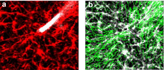
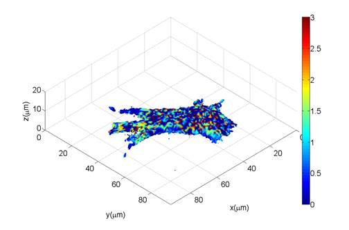
We used live-cell confocal imaging to simultaneously track cell migration and fibrin matrix distortion by human foreskin fibroblasts (HFFs) on timescales ranging from seconds to hours. Consistent with previous studies of fibroblastic cell types, HFFs generate spindle-like protrusions and contract the fibrin matrix in a dynamic manner. Our preliminary data suggest that myosin localization in the protrusion precedes local contraction of the matrix, and that protrusions deform the matrix in successive contract and release cycles, consistent with local mechanical feedback (Figure 1). Our data additionally yield the surprising observation that changes in cell volume correlate with global deformations of the surrounding matrix: 60% of cells (n = 18) underwent 10% or greater changes in volume in ~ 3 hours, and both increases and decreases in cell volume coincided with matrix deformation (Figure 2). Regulated changes in cell volume may thus constitute a presently overlooked mechanism for cell-driven matrix remodeling in 3D matrices and tissues. Ongoing work examines the role of mechanically activated signal transduction pathways in mediating matrix deformation, both at cell protrusions and in conjunction with cell volume regulation.
|
|
Figure 1. Matrix deformations in a fibrin gel due to protrusive activity of a fibroblast. (a) A fibroblast protrusion (GFP actin) in a fibrin matrix (red). (b) Vectors showing the matrix deformation. (image size: 33 x 29 µm) | |
|
Figure 2. Matrix deformation due to volume change of a fibroblast embedded in a 3D fibrin matrix. The volume of the cell decreased by 10% over 90 minutes. The 3D deformations of the fibrin matrix were quantified using digital volume correlation (DVC) and mapped on the cell surface. Heat map shows the displacement of the matrix in microns. |
See more of this Group/Topical: Food, Pharmaceutical & Bioengineering Division