
 Google
 Yelp
 Facebook
 Twitter
 ChEnected
334108 Nanostructured 3D PLGA Scaffolds For Skin-Tissue Engineering Applications
NANOSTRUCTURED 3D PLGA SCAFFOLDS FOR SKIN-TISSUE ENGINEERING APPLICATIONS
Z. Karahaliloğlu1, B. Ercan2, E.B. Denkbaş3, T. Webster2
1Hacettepe University, Nanotechnology and Nanomedicine Division, 06800, Beytepe, ANKARA, Turkey
2Northeastern University, Chemical Engineering Department, 02115, Boston, MA, USA
3Hacettepe University, Chemistry Department, Biochemistry Division, 06800, Beytepe, ANKARA, Turkey
Background and aims: Skin is the largest organ in the body and performs various functions (i.e., maintaining temperature, waste exchange, etc.) critical for our survival. When skin-tissue is damaged due to a burn or a trauma, natural skin grafts in the form of a xenograft, allograft or autograft have been providing optimal results for wound healing applications. However, the use of graft tissue comes with numerous challenges including limited donor site for severe skin trauma patients, potential for immune response for allographs and xenographs, requirement of a surgical procedure, and high cost1,2. Although some of the commercially available synthetic skin substitutes regenerate some of the functions of damaged epidermal tissue, there is no currently-used material that can replace the functional complexity of natural skin. To address this challenge, we are proposing the use of nanofeatured poly(lactic-co-glycolic acid) (PLGA) as an alternative skin substitute. PLGA is an FDA approved biodegradable and a biocompatible copolymer used in various medical applications such as resorbable sutures, surgical clips and drug delivery systems3. Furthermore, there is plenty of in vitro and vivo study showing that nanostructured PLGA enhances cellular adhesion, proliferation and long-term functions4,5. In this research study, nanostructured 3D PLGA scaffolds were created and investigated as an alternative material for skin-tissue engineering and its regenerative performance was compared to a nanosmooth PLGA scaffold in vitro.
Methods: In this work, PLGA scaffolds were prepared by solvent casting and salt leaching process as reported previously4. Briefly, 0.5 g of PLGA (PLGA; 50:50 wt%; Polysciences, Inc.) were dissolved in 6 ml of chloroform in 45 °C for 30 min. 4.5 g of sodium chloride salt (NaCl, up to 250 µm, Sigma) were used to introduce pores into the substrate. NaCl particles were added to the dissolved polymer solution. After mixing and sonicating for 20 min, the mixture was poured into a Teflon petri dish and dried overnight at room temperature. To evaporate of all chloroform, the dried polymer was placed into a vacuum for 48 h. The scaffold was soaked in deionized water for 3 days. To understand the effect of PLGA nanorough surfaces on human epidermal keratinocyte (İnvitrogen C0015C) and fibroblast cell lines (ATCC CCL-110), PLGA scaffolds were soaked in a NaOH solution prepared at different concentrations (0.1, 0.5 and 1 N) and for various times (5, 10, 15 and 20 minutes). Characterization of the treated- and untreated PLGA scaffolds was performed by using Scanning Electron Microscopy (SEM). Cell adhesion to the scaffold and cell proliferation was assessed using human fibroblast and human keratinocyte cell lines. Experiments were completed in triplicate and repeated at least three times.
Results: SEM images of untreated and treated PLGA scaffolds exhibited a significant change in surface roughness. Treated PLGA had nano topographical surface properties while untreated PLGA scaffolds showed a smooth surface. Our preliminary results (not shown here) indicate that nanofeatured PLGA surfaces promoted more skin cell adhesion compared to its nanosmooth counterpart.
Conclusion: This study confirmed that PLGA scaffolds altered to have a nanorough surface topography by chemical etching technique should be further studied for improved skin-tissue engineering applications.
| a |
| b |
| d |
| c |
| e |
| f |
| g |
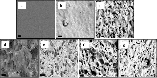
Figure 1. SEM images of (a) untreated and (b) 0.1 N NaOH-treated for 10 min, (c) 0.5 N NaOH-treated for 10 min, (d) 1 N NaOH-treated for 5 min, (e) 1 N NaOH-treated for 10 min, (f) 1 N NaOH-treated for 15 min and (g) 1 N NaOH-treated for 20 min PLGA scaffolds. Scale bars= 200 nm.
Acknowledgements: The authors would like to thank Daniel Hickey and Linlin Sun for their assistance. The authors would also like to thank Northeastern University and Hacettepe University for funding.
References
1. Sheila, M. Nature, 2007, 445, 874¨C880.
2. Marler, J. J.; Upton, J.; Langer, R.; Vacanti, J. P. Advance Drug Delivery Rev. 1998, 33, 165-182.
3. Kumbar, S.G.; Nukavarapu, S.P.; James, R;, Nair, L.S.; Laurencin, C.T. Biomaterials, 2008, 29, 4100¨C4107.
4. Smith, L.L.; Niziolek, P.J.; Haberstroh, K.M.; Nauman, E.A.; Webster, T.J. Int J Nanomedicine, 2007, 2(3), 383¨C388.
5. Park, G.E.; Pattison, M.A.; Park, K.; Webster, T.J. Biomaterials, 2005, 26(16), 3075.
See more of this Group/Topical: Nanoscale Science and Engineering Forum