
 Google
 Yelp
 Facebook
 Twitter
 ChEnected
336058 The Direct Osteolytic Potential of MCF-7 and MDA-MB-231 Breast Cancer Cells in An in Vitro Bone Environment
The direct osteolytic potential of MCF-7 and MDA-MB-231 breast cancer cells in an in vitro bone environment
JE Dumas, CJ Ihenacho, and MO Platt
Department of Biomedical Engineering, Georgia Institute of Technology, Atlanta, GA
Introduction. Breast cancer affects one in eight women, and its propensity to metastasize to bone causes further health complications. Cathepsins and other proteases have been reported to be upregulated in breast cancer tissues, facilitating the invasion and metastasis of breast cancer. It is generally established that cancer cells influence osteoclast activity by releasing factors such as parathyroid hormone that stimulate the production of RANKL, which increases osteoclastogenesis and increased bone resorption (i.e., bone degradation). Growth factors released from the degraded matrix then bind to cancer cells stimulating proliferation. This vicious cycle causes osteolytic lesions and considerable patient morbidity. The direct resorptive capacity of breast cancer cells is not well understood and could contribute to this osteolytic cycle. Therefore, it is important to fully elicit putative mechanisms of direct resorption of bone by metastasized breast cancer cells to optimize bone metastasis treatment strategies. To do so, engineered bone biocomposites, are useful to determine individual mechanisms behind these pathologies in a controlled environment. In this study, a bone biocomposite platform is used to study the induction of bone resorptive activity by two breast cancer cell lines noted for differences in their aggressive/metastatic abilities MDA-MB-231, and MCF-7.
Materials and Methods. Bone biocomposites were synthesized using polyurethane and bovine mineralized bone particles. The bone biocomposites were molded in cylindrical rods, and were subsequently cut into discs (500 µm thickness). After sterilization, the discs were placed in a 12 well plate and 5.0 x 10⁴ MDA-MB-231and MCF-7 cells (transfected with RFP and GFP, respectively) were seeded on the composite surface. Media was collected every two days, and cells were lysed after seven days. Protein concentration was determined and lysates were prepared for Western blot. Media was analyzed for calcium using an Arsenazo III assay. Collagen fragment (CTX) concentrations in cell media were measured using ELISA.
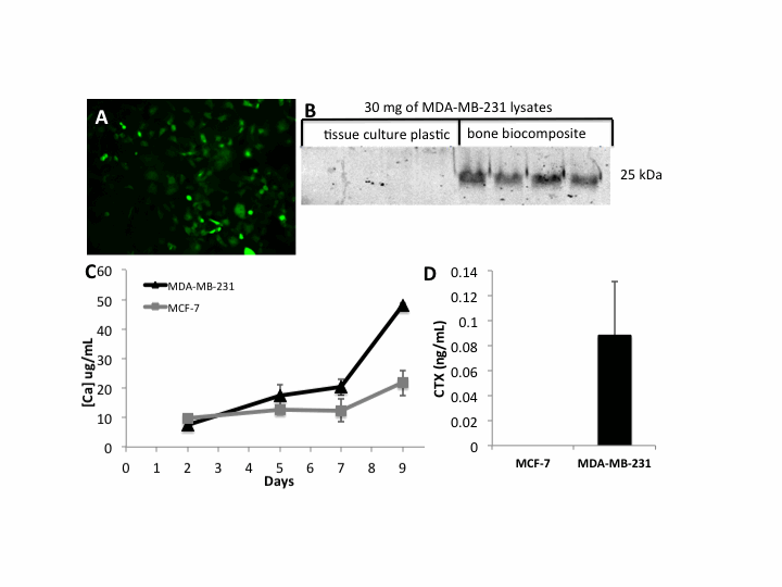
Results and Discussion. After nine days of culture, MDA-MB-231 exhibited a 2-fold increase in calcium released into the medium as compared to MCF-7s and all above the background no cell control. Increased calcium released in the culture media was an indicator that breast cancer cells, in the absence of active osteoclasts, may be capable of resorbing bone, and that the bone biocomposites we developed may be a useful tissue engineered platform to study these mechanisms. Further, culture on the bone biocomposites but not on tissue culture plastic, exhibited increased expression of cathepsin K, the major enzyme that degrades type I collagen (the major structural component of bone), which is coupled with bone de-mineralization. Increased cathepsin K expression coincides with increased CTX concentrations in the culture media.
Conclusions. Bone biocomposites synthesized in this study demonstrated that breast cancer cells are capable of mineral dissolution and collagen cleavage that may lead to osteolytic lesions in metastasized cancer. This observation emphasizes the importance of early intervention of bone metastasis as the breast cancer cells may directly begin resorption of bone, prior to activation of the neighboring osteoclasts and may present a mechanism that improves their homing and colonization of the foreign environment.
Figure SEQ Figure \* ARABIC 1. (A) Fluorescent image of MCF-7 cells attached on the surface of a bone biocomposite at day 4. (B) Western blot indicating an increase of cathepsin K expression of MDA-MB-231 cells seeded on bone biocomposite as compared to cells seeded on tissue culture plastic. (C) Calcium accumulated in culture media from bone biocomposite degradation over a 9 day period of both MDA-MB-231 and MCF-7 cells. (D) CTX collagen fragments accumulated in culture media between days 7 and 9.
See more of this Group/Topical: Materials Engineering and Sciences Division