
 Google
 Yelp
 Facebook
 Twitter
 ChEnected
336222 Force-Dependent Interactions Between Actin Filaments and a Minimal Adherens Junction Complex
Introduction
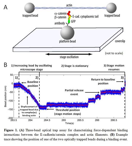
Adherens junctions (AJs) mediate adhesion between neighboring cells and are essential for coordinating cell-cell adhesion and cytoskeletal dynamics. Disruption of cytoskeletally generated tension in living cells impairs AJ formation, suggesting that localized mechanical stress may be required for AJ assembly. Previous attempts to measure the interaction of the core AJ components E-cadherin, beta-catenin, and alpha-catenin with actin in vitro have been unsuccessful, perhaps due to the absence of a mechanical load in bulk biochemical assays. Here we describe a new optical trap-based single-molecule assay that allows us to directly probe the interaction of the E-cadherin/catenin complex with filamentous actin as a function of applied mechanical load (Fig. 1A). Optical traps (also known as optical tweezers) utilize highly focused laser light to capture and exert force on micron-sized dielectric objects. The instrument we have constructed consists of a dual-beam optical trap that can apply up to ~20 piconewtons (pN) of force while measuring the location of a trapped 1 mm polystyrene bead with nanometer and millisecond resolutions.
Results
Our assay works as follows: two streptavidin-coated polystyrene beads are trapped simultaneously using the dual-bead optical trap and a biotinylated actin filament is stretched between them. This filament is then lowered over a platform bead immobilized on a coverslip and functionalized with the reconstituted E-cadherin/beta-catenin/alpha-catenin complex. Oscillation of the piezo-actuated microscope stage moves the platform bead and attached E-cadherin/catenin complexes back and forth along the axis of the actin filament while the optical traps remain stationary. Load is applied when the immobilized complexes bind to the filament as the stage continues moving and one of the two beads is pulled away from the trap center. As the bead is pulled away, the force gradient produced by the focused laser beam attempts to pull the bead back to the center of the optical trap. The farther the bead is pulled, the greater the restoring force it and the attached protein complex experience. The microscope stage velocity and the stiffness of the optical traps thus determine the force loading rate experienced by the E-cadherin/catenin complexes. Using high concentrations of the E-cadherin/catenin complex (~10 to 100 nM), we observe binding events that likely involve the attachment of multiple complexes to a single actin filament. Under these conditions, we observe displacement events of over 100 nm with loading rates of ~45 pN/s that can sustain over 15 pN of force. Detachment occurs via a stepwise release mechanism that may reflect sequential detachment of individual complexes (Fig. 1B).
Conclusions
Our data indicate that several E-cadherin/catenin complexes can together form a robust, load-resistant connection to an actin filament under tension. This observation is consistent with a hypothesized role for alpha-catenin in linking the adherens junction to the actin cytoskeleton. Ongoing work tests the possibility that the E-cadherin/catenin complex acts as a mechanosensitive element whose affinity for actin is directly modulated by mechanical load.
See more of this Group/Topical: Food, Pharmaceutical & Bioengineering Division