
 Google
 Yelp
 Facebook
 Twitter
 ChEnected
337618 3-Dimensional Osteochondral Tissue Engineering
Osteoarthritis affects millions of individuals and is characterized by joint pain caused by articular cartilage wear. Cartilage, due to its avascular nature, has poor self-repair capabilities, which result in mechanically inferior repair tissue. One of the more successful current clinical techniques involves replacing the damaged cartilage and part of the underlying subchondral bone with osteochondral grafts. However, the availability of such grafts is limited and if taken from the patient, results in another defect site. Therefore, tissue engineering has been investigated as a promising solution for cartilage defects. In this study, we developed a novel gradient generating bioreactor that was used to investigate tissue engineering of osteochondral tissues by using molecular gradients to control human mesenchymal cell (MSC) differentiation. Within the bioreactor, a flow channel with two inlets allows for the flow of molecular gradients across 3-dimensional tissue constructs (Fig. 1 and Fig. 2). Using computational fluid dynamics simulations, we designed the reactor and showed that these flow established gradients can be created and maintained indefinitely (Fig. 2). Further, through fluorescent tracer experiments, we demonstrated the generation and maintenance of the gradients of bioactive molecules (Fig. 3). To test the effect of molecular gradients on chondrogenesis, we exposed human MSCs within 3-dimensional collagen scaffolds to gradients of transforming growth factor beta 1 (TGF-β1). Chondrogenesis was visualized via toluidine blue staining for glycosaminoglycans and was only observed within the construct regions exposed to higher TGF- β1 concentrations (Fig. 4). To develop osteochondral constructs, the tissue constructs were exposed to opposing gradients of chondrogenic and osteogenic media. The bioreactor designed here allows for the production of stable molecular gradients that can be controlled by adjusting various parameters including the inlet flow rates, inlet molecular concentrations, and tissue construct position within the reactor (distance from inlets). The reactor can also serve as a platform to study the effect of molecular gradients on other composite tissue constructs.
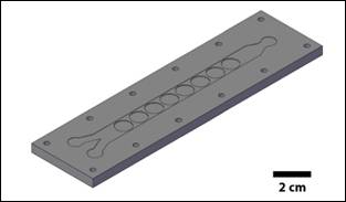
Figure 1: Bioreactor Design. Two inlets combine into a single flow channel to form a continuous molecular gradient across the width of the channel. Three dimensional tissue constructs are placed inside circular wells below the flow channel.
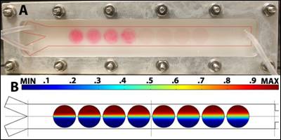
Figure 2: Bioreactor and Gradient Simulation. A. Assembled bioreactor containing MSC-collagen sponge constructs within the first 4 wells. B. Gradient simulation across each of the wells of the bioreactor.
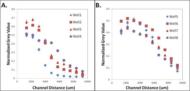
Figure 3: Fluorescent Gradient Characterization. Fluorescent intensity (normalized grey value) was measured across the width of the flow channel at each well containing collagen sponges. Wells are numbered starting from the well closest to the inlets (well 1). A. Wells 1-4. B. Wells 5-8.
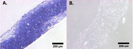
Figure 4: Chondrogenesis of MSC-Collagen Sponge Constructs. Toluidine blue staining for MSC-collagen sponge constructs exposed to A. higher TGF-β1 concentrations and B. lower TGF-β1 concentrations along the gradient.
See more of this Group/Topical: Food, Pharmaceutical & Bioengineering Division