
 Google
 Yelp
 Facebook
 Twitter
 ChEnected
337650 Reduced Adhesion of Macrophages On Nanotubular PDMS Molds for Catheter Applications
Introduction: Polydimethylsiloxane (PDMS) has been used for short- and long-term catheters. However, PDMS shunt tubing has been scrutinized recently because of their extremely high failure rates. While there are many reasons why PDMS shunt systems fail, one is tissue occlusion of the ventricular catheter, most often caused by macrophages. It is hypothesized that nanotextured and nanotubular surfaces can be carefully manipulated to inhibit immune cell (specifically, macrophages) and improve the fibroblast adhesion due to their unique surface energy properties which have the ability to control initial protein absorption and subsequent cell behavior. The objective of this in vitro study was to create polydimethylsiloxane (PDMS) molds of titanium (Ti) anodized to possess nanotubes and test inflammatory cell and fibroblast responses on such substrates.
Materials and Methods: 99.2% pure titanium foils (Alfa Aesar, Ward Hills, MA) were cut into 1cm×1cm squares and cleaned with acetone, 70% ethanol, deionized water. The samples were etched for 1min with a solution of 1.5% nitric acid and 1.5% hydrofluoric acid (HF) to remove the thin oxidized layer. The cleaned Ti sample was used as an anode, while a platinum (Pt) mesh served as a cathode. Both were immersed in an electrolyte solution consisting of 1.5% HF. The Ti and Pt samples were connected to a DC power supply. A constant voltage of 20V was applied for 10 minutes and the distance between Ti anode and Pt cathode was kept constant at 1cm. After anodization Ti samples were rinsed with large amounts of deionized water immediately and air dried.
Next, the PDMS monomer and cross-linking agents (Sylgard 184, Dow Chemical Co.) were mixed at 10:1 (weight ratios) for 15min and were then placed in a vacuum chamber for 30min to remove air bubbles. The mixture was cast onto the nanotubular Ti master mold and then was placed into a vacuum chamber for another 1h. The PDMS was cured at 60 °C for 2h followed by cooling and was gently peeled away from the Ti template. To improve the peeling off ability, a gold film was sputtered onto the top surface of the PDMS template and the PDMS slurry was again poured onto the first PDMS. The desired PDMS replica was obtained after curing.
The IC-21 macrophage cell line was used (TIB-186; ATCC, Manassas, VA) for determining an immune response. Macrophages were cultured according to ATCC instructions in RPMI-1640 medium (Invitrogen, Carlsbad, CA) with 10% fetal bovine serum and 1% penicillin/streptomycin (HyClone Laboratories Inc, Logan, UT) under standard incubator conditions (37°C, humidified, 5% CO2/95% air environment). Macrophages were seeded at 3500 cells/cm2 per substrate and were cultured in a humidified 5% CO2/95% air environment at 37°C for 24 hours. After 24 hours, nonadherent macrophages were removed by rinsing in phosphate-buffered saline (PBS). Macrophages adherent on the substrates were fixed with 4% formalin in PBS and stained with both rhodamine phalloidin (R415) (Molecular Probes, Eugene, OR) and DAPI (4', 6-diamidino-2-phenylindole) (33258) (Sigma-Aldrich). F-actin filaments and cell nuclei were visualized using tetramethyl rhodamine iso-thiocyanate and DAPI, respectively.
Also, to determine the adhesion and proliferation of skin fibroblasts (ATCC, CCL-110), a cell proliferation assay (CellTiter 96, Promega) was used. Briefly, for cell adhesion, cells were seeded at 3500 cells/cm2 in standard cell culture media and were incubated for 4 hours. For proliferation studies, cells were seeded at 3500 cells/cm2 for 1, 3 and 5 days. The dye solution were added to the cells after the end of the prescribed period for 4h, then the stop solution was added and incubated overnight. A plate reader was used to test cell density. Numerical data were analyzed with a Student's t-test to make pair-wise comparisons. Statistical significance was considered at p<0.05.
Results and Discussion: As expected, compared with unmodified Ti (Figure 1. a), nano-sized tubes were distributed uniformly on the Ti surface after anodization (Figure 1, c). The uniform pores, as observed by scanning electron microscopy, were estimated to have a diameter of 60-70nm and a depth of 200nm. After pouring the PDMS slurry onto the surface of the nanotubular Ti and peeling off the first PDMS template, a nanopatterned structure was observed (Figure 1, d). Most importantly, the PDMS replica showed similar nanostructures as anodized Ti.
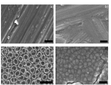
Figure 1. SEM images of
a) Unanodized Ti b) PDMS based on unanodized Ti
c) Anodized Ti d) PDMS based on anodized Ti
Scale bars are 200nm.
The results also demonstrated decreased numbers of macrophages adhered to nanotubular Ti and PDMS replica surfaces. Most importantly, the increased fibroblast adhesion, proliferation, and long-term functions on nanotubular substrates indicated the nanoscale topographies of polymers should be strongly considered for catheter applications.
Conclusions: A PDMS replica was obtained from anodized nanotubular Ti and demonstrated a promising ability to control macrophage and fibroblast functions.
Acknowledgements: The authors would like to thank Northeastern University for funding.
See more of this Group/Topical: Nanoscale Science and Engineering Forum