
 Google
 Yelp
 Facebook
 Twitter
 ChEnected
337655 The Role of Actomyosin Generated Tension in Coordinating Cell Movements During Zebrafish Development
Introduction
Cells engage in collective migration in circumstances ranging from wound healing to embryonic development. While much is known about how single cells migrate, it is unclear how groups of cells coordinate movements across large distances. In this study we use zebrafish embryos as a model system to investigate the role of cell-generated line tensions in generating long-range, coordinated motion. Early zebrafish development exhibits intricately orchestrated cell migration and patterning. Within the first 24 hours a mass of cells termed the blastoderm spreads uniformly over a spherical yolk, starting at one end and moving toward the opposite pole in a process termed epiboly. During epiboly, the cells at the blastoderm margin (edge) migrate at a uniform rate and thus reach the opposite pole of the yolk at the same time (Figure 1A-C). This long-range coordination is essential in establishing the axes that define the front, back, top and bottom of the resulting organism. A band of actin and myosin, termed the actin band, forms near the blastoderm margin. It has been previously shown that the actin band produces a contractile tension; however its functional role is not known.
Results
We used mechanical shape perturbations to probe the function of the actin band during epiboly. In shape-perturbed embryos, we find that the rate of blastoderm migration is no longer uniform: lagging areas of the margin speed up, while leading areas slow down. This equalizes progress along the blastoderm margin perimeter such that epiboly reorients to coincide with the long axis of the embryo. When the tension within the actin band is disrupted using chemical treatments such as blebbistatin or reduction of calcium this reorientation no longer occurs. This observation suggests that the tension within the actin band is a necessary component in coordinating blastoderm migration along the length of the margin. We additionally observed that cells along the blastoderm margin align parallel to the actin band. Treatment with either blebbistatin or calcium depletion resulted in a markedly rougher margin, suggesting that the actin band additionally coordinates motion between neighboring cells.
Discussion
Based upon these results, we developed an analytical model in which the actin band acts like a pre-tensioned rubber band, introducing a line tension that coordinates blastoderm migration over millimeter length scales (Figure 1D). This line tension also provides a resistive force against instabilities at the margin of the migrating blastoderm, ensuring that neighboring cells progress at similar rates (Figure 1E). Our model is in quantitative agreement with margin migration rates observed in a wide variety of embryo shapes, suggesting that a simple physical parameter (actin band tension) is sufficient to account for the long-range cell coordination that we observe. The physical properties of the zebrafish blastoderm margin are similar to those of spreading cell sheets observed in a wide variety of circumstances. We suggest that similar quantitative, physical models may account for collective cell movements observed in a wide variety of circumstances including other developmental processes, wound healing, and a variety of tissue engineering applications.
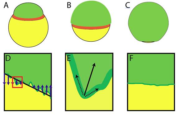
Figure 1. Mechanical tension leads to long-range coordination between blastoderm cells in the zebrafish embryo. A-C) Progression of epiboly (green – blastoderm, yellow – yolk, orange – actin band). A) Epiboly begins with the blastoderm sitting on one pole of the yolk cell. B) The blastoderm migrates over the yolk cell until it finishes epiboly by converging at the pole opposite the initial blastoderm pole (C). D-F) Description of the roles of actomyosin generated line tension. D) Line tension (black line) along the boundary of a migrating cell mass (green) generates local forces (blue) that reorient the cell boundary. E) Zoomed in view of an instability at the boundary (red square in D). Tension at the margin (black dotted arrows) generates a net restorative force (black solid arrow). F) As a result, the cell mass reorients and the boundary maintains a smooth profile that is parallel to the actin band.
See more of this Group/Topical: Food, Pharmaceutical & Bioengineering Division