
 Google
 Yelp
 Facebook
 Twitter
 ChEnected
340110 Increased Osteoblast Adhesion and Proliferation On Electrophoretic Deposition Coated Nano Hydroxyapatite On Titanium-6,4
INCREASED OSTEOBLAST ADHESION AND PROLIFERATION ON ELECTROPHORETIC DEPOSITION COATED NANO HYDROXYAPATITE ON TITANIUM-6,4
Garima Bhardwaj1, Dennis Mathew1, Linlin Sun2, Thomas J.Webster2 and Geetha Manivasagam3.
1Department of Biomedical Engineering, VIT University,Vellore-632014,Tamil Nadu,India.
2Department of Chemical Engineering, Northeastern University, Boston, MA-02115, USA.
3Department of Mechanical Engineering, VIT University,Vellore-632014,Tamil Nadu,India.
Introduction:Improving the performance of titanium implants for orthopedic applications has been a concern for many years.This can be completed by designing surface properties to promote osseointegration. One manner to improve orthopedic implant performance is to coat metals with bioactive ceramics, such as nanocrystalline hydroxyapatite, which have been shown to improve bone formation compared to micron crystalline hydroxyapatite and other ceramics. However, current hydroxyapatite coating methods are time consuming, may be expensive, and involve high heat which transforms nanocrystalline hydroxyapatite into micron crystalline hydroxyapatite decreasing bioactivity and decreasing coating adhesion strength.The goal of this present research was to develop a novel, quick, and inexpensive implant coating process to improve bone cell responses.
Materials and Methods:All of the tests in this study were performed on a titanium alloy, Ti-6Al-4V, which was cut into small pieces with dimensions of 1cm X 1 cm. Nanophase hydroxyapatite (HAP) was prepared by wet chemical synthesis using Ca (NO3)24H2O, KH2PO4,distilled water, ammonia and acetone with a stirring time of 1 hour and an aging time of 24 hours followed by filtering then sintering at 900 ͦC. The HAP was then coated onto the alloy using electrophoretic deposition (EPD). The electrolyte was prepared by dissolving HAP in isopropyl alcohol and stirring for 10 minutes followed by overnight settling. The mixture was again stirred for 2 hours before starting the coating and Ti-6,4 was used as a working electrode(-ve) and 316 stainless steel was used as a counter electrode(+ve).Voltage varying from 40 to 80 V was used for 2 to 5 minutes and an optimal coating was achieved at 60 V for 4 minutes. Samples were characterized using X-ray Diffraction (XRD), SEM, and contact angle analysis to confirm crystallinity, surface roughness, and chemistry of the coatings. Osteoblasts (ATCC-C12720) were cultured in growth medium supplemented with 1% penicillin.A 4 hour MTT adhesion assay was performed to determine cell viability.A MTT proliferation assay was performed after 1,3 and 5 days of culture.All experiments were conducted in triplicate and repeated three times each.
Results and Discussion XRD results of the nanophase HAP (Figure 1) and SEM images of the samples taken after the coating process (Figure 2) confirmed the uniformity and nanoscale character of the coatings. Contact angle results of the coated samples showed an increased hydrophilic nature of the coated samples. Results from the MTT assay showed increased osteoblast adhesion (Figure 3) and proliferation (Figure 4) on the coated samples compared to controls (tissue culture polystyrene and uncoated Ti-6Al-4V).
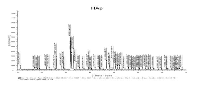
Figure 1: XRD of nanocrystalline HA used in the present study
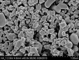
Figure 2. SEM of the EPD nanocrystalline HA on titanium-6,4
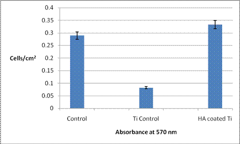
Figure 3. Increased osteoblast adhesion on HA (EPD) coated Ti compared to Ti control. Data = mean +/- SEM; n = 3 * p < 0.01 compared to Ti control,F-test value=0.000356.
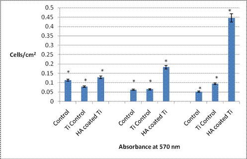
Figure 4: Osteoblasts proliferation results, day 1, 3 and 5 respectively.Data = mean +/- SEM; n = 3; * p < 0.01 compared to Ti control,F value for Day 1=0.001872,F value for Day 3=0.0017608,F value for Day 5=0.007834
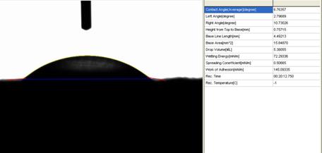
Figure 5:Contact angle analysis results(actual picture and values of drop and surface contact) for uncoated Ti-6,4 obtained using distilled water as a fluid.
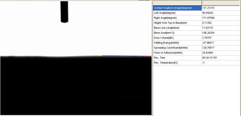
Figure 6:Contact angle analysis results(actual picture and values of drop and surface contact) for Ti-6,4 coated with nano-HA by EPD obtained using distilled water as a fluid.
Conclusions: Electrophoretic deposition is a novel, inexpensive and quick coating process that leads to a strong and optimal coating to increase bone cell compatibility. Hence, this coating process should be further explored for enhancing orthopedic implant efficacy.
Acknowledgements: The authors thank Northeastern University and VIT University for funding.
See more of this Group/Topical: Nanoscale Science and Engineering Forum