
 Google
 Yelp
 Facebook
 Twitter
 ChEnected
340736 An Injection-Molded Device for Purification of Nucleic Acids From Whole Blood Using Isotachophoresis
An Injection-Molded Device for Purification of
Nucleic Acids from Whole Blood using Isotachophoresis
Lewis A. Marshall, Anita Rogacs, Carl Meinhart, and Juan G. Santiago
Stanford University
Stanford, CA 94305, USA
We have designed a new microfluidic chip to simplify nucleic acid preparation from biological samples such as whole blood. We previously demonstrated the application of isotachophoresis (ITP) for sample preparation.1 We here describe the design of an injection molded plastic device capable of purifying DNA from a 25 µL sample of whole blood lysate under 40 min.
We have targeted samples with volumes between 2-25 µL in an attempt to address an unmet need in sample preparation. Solid phase extraction (SPE) kits for purification of nucleic acids work well for sample volumes of 100-1000 µL, and nucleic acid yields of several nanograms. However, they exhibit poor extraction efficiency for samples of low volume and smaller nucleic acid yields. ITP does not rely on surface adsorption, and so can operate on samples with a wide range of volumes and nucleic acid content. Our new device enables ITP purification of samples in this critical volume range while simplifying device operation. We have also maximized device throughput by adjusting our chemistry and protocol and considering the sample volume processing rate as a cost function..
This new device is pictured in Figure 1. This custom device is fabricated from injection molded Topas cyclic olefin copolymer (COC) or poly(methyl methacrylate) (PMMA). The device was fabricated by the microfluidics foundry Microfluidic Chipshop GmbH of Jenna, Germany. The injection molded layer contains channels with a nominal cross-section of 2 mm x 150 µm, a total length of 16 cm. The injection molded layer is capped by a thin, transparent plastic film to enable imaging into the channel.
We've included novel features in this chip to improve the device loading process. To simplify set up of the initial interface between the sample and the separation buffer, we've developed a specialized capillary barrier structure that provides passive valving for sequential loading of liquids into the device. This structure is shown in Figure 2. In addition to incorporating this passive valving system for device loading, we have incorporated the use of gels in the buffering reservoirs to suppress pressure driven flow. This simplifies loading by making the device performance robust to uneven fluid levels in the reservoirs.
We used this device to extract DNA from whole blood lysate samples. A visualization of the separation process is shown in Figure 3. Here, we separate two fluorescent dyes with mobilities similar to DNA and a contaminating protein species. The analyte dye focuses into a sharp band and elutes into the extraction reservoir, while the contaminating species lags behind and within the channel for later disposal.
We verified the purity of our extracted samples from species that inhibit polymerase chain reaction (PCR) by performing quantitative PCR (qPCR) and comparing the results with those collected from samples extracted using a commercial SPE DNA purification kit. Sample qPCR data is shown in Figure 4.
By introducing a standard injection molded device for purification of samples using ITP, we have created a method of performing sample preparation at a relevant scale using ITP. This system addresses an unmet need in sample preparation of nucleic acids by providing an alternative method for processing samples with low nucleic acid content.
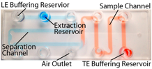
Figure 1. Image of the new injection molded device. For imaging, the device was filled with colored liquid to improve contrast. The sample channel (red) can hold a 25 µL sample. The separation channel (blue) holds 28 µL separation buffer. There are buffering reservoirs for both the leading and trailing electrolyte (LE and TE). These buffering reservoirs contain a gel with high buffering capacity gel to maintain a constant pH in the channel while suppressing pressure-driven flow. An air outlet with specialized capillary structures is included to ease the loading process, as shown in Figure 2.
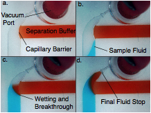
Figure
2. Capillary loading structures near the vacuum
port. The capillary structures allow set-up of a fluid interface without
wasting sample or trapping bubbles. a. The separation channel is initially filled
with the (red) separation buffer, which wets up to the surface of the capillary
barrier. b. The sample is applied to the sample channel, and a -0.1 psig vacuum is
applied to the air outlet to draw the sample into the channel. c. The sample
wets the separation buffer, causing it to break through the barrier. d. The liquids
form a sharp interface, and flow up to the final fluid stop barrier.
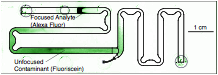
Figure 3. An experimental image of separation of two dyes overlaid with the device schematic. Two dyes, Alexa Fluor 488 (AF) and fluorescein (FL) are loaded as the sample. When current is applied, the AF focuses to the ITP interface, while the FL remains behind as an unfocused zone. The AF is about to elute into the extraction reservoir, while the FL remains behind in the channel.
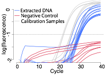
Figure 4. qPCR results. We performed qPCR with samples extracted from whole blood using isotachophoresis. The extracted DNA amplified consistently, and at the predicted temperature for the amplified sequence (not shown). The results were comparable to qPCR amplification of calibration samples with template DNA extracted from blood using a commercially available SPE kit. We performed negative controls using whole blood lysate, water, and the separation buffer as templates, and observed no amplification.
REFERENCES:
1. "Purification of nucleic acids from whole blood using isotachophoresis," Persat, A., Marshall, L. A., and Santiago, J. G. Analytical Chemistry 81, 9507 (2009).
See more of this Group/Topical: 2013 Annual Meeting of the American Electrophoresis Society (AES)