
 Google
 Yelp
 Facebook
 Twitter
 ChEnected
341191 Endotoxin Binding By the Cationic Amphiphilic Peptide WLBU2 in Relation to Polymyxin B
Introduction: Sepsis is a blood infection that in the US alone affects about 750,000 people each year, killing 28-50% of them.1-3 Passing blood through a sorbent device (hemoperfusion) to specifically remove targets such as endotoxin and bacterial cells holds promise for rapid treatment of acute sepsis. Clinical use of hemoperfusion in this context is based on immobilized polymyxin B (PMB),4,5 but remains limited owing to ineffective endotoxin removal, and serious complications (e.g., nephrotoxicity, neurotoxicity, monocyte stimulation, substantial protein loss) associated with PMB and PMB-based devices4,6-9. The synthetic, cationic amphiphilic peptide (CAP) WLBU2 has greater antimicrobial activity than PMB, works against a much broader spectrum of Gram-positive and Gram-negative bacteria, and shows higher selectivity for pathogenic entities over host cells.10,11 In this research WLBU2 was compared to PMB with respect to its ability to bind endotoxin in the presence and absence of blood proteins, while in solution or immobilized at surfaces. Endotoxin binding of WLBU2 while covalently tethered to a surface was also investigated.
Materials and Methods: Optical waveguide lightmode spectroscopy (OWLS) was used to detect adsorption on hydrophilic and silanized (hydrophobic) SiO2 coated OW2400c waveguides under steady flow conditions. Vapor-phase silanization was carried out with trichlorovinylsilane (TCVS). Peptide (WLBU2 or PMB) and endotoxin (lipopolysaccharide, LPS, isolated from Pseudomonas aeruginosa), in PBS or diluted human plasma, were introduced to the OWLS flow cell in sequence or in mixed suspensions. Each adsorption step was followed by an elution step in peptide-free, LPS-free PBS buffer. LPS vesicle adsorption and spreading behavior under conditions representative of the OWLS experiments were also recorded, using interfacial tensiometry.
Results and Discussion: The antimicrobial effect of PMB and many CAPs is attributed to membrane disruption in susceptible bacteria. Assuming a multi-step mechanism for binding and disruption, our results strongly indicate that in contrast to PMB, WLBU2 binding is not accompanied by rapid disruption of LPS bilayer vesicles. In particular, competitive adsorption results showed adsorption of WLBU2 to hydrophobic surfaces from peptide-LPS mixtures is reduced by an order of magnitude relative to WLBU2 in the absence of LPS. Peptide-LPS and LPS-peptide sequential adsorption results were consistent with this outcome. Figure 1 shows that after LPS layering at a hydrophobic surface, WLBU2 binding was entirely irreversible with respect to buffer dilution.
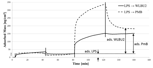
Figure 1: Sequential adsorption of LPS and WLBU2 (solid line) or PMB (dashed line). After introduction of peptide, the resistance to elution by PBS is much more pronounced in the case of WLBU2, indicating peptide “entrapment” within the LPS layer. This is consistent with binding affinity of WLBU2 for LPS.
Conclusions: Over the timescales of these experiments, we have seen that WLBU2 tightly binds LPS in a fashion which leaves the LPS largely intact. In contrast, PMB binding to LPS is associated with vesicle disruption and evolution of disruption products. Use of WLBU2 to capture pathogenic entities in a hemoperfusion device would avoid adverse clinical effects due to reintroduction of such entities to circulating blood. These early investigations of use of tethered WLBU2 to capture LPS are promising. Further investigation will be conducted using tethered constructs in the presence of blood proteins, as well as against whole bacterial cells.
References
8. Ikeda T. Hemoadsorption in critical care. Therapeutic Apheresis 2002;6(3):189-192.
See more of this Group/Topical: Materials Engineering and Sciences Division