
 Google
 Yelp
 Facebook
 Twitter
 ChEnected
341618 Metabolomic and Proteomic Analysis of Metabolic Enzyme Disruption in Adipocytes
Metabolomic and Proteomic Analysis of Metabolic Enzyme Disruption in Adipocytes
Introduction: Obesity results from a chronic imbalance in caloric intake and expenditure. The excess calories are stored mainly as intracellular lipids (triglycerides, TG), leading to an expansion of body fat or adipose tissue through increases in fat cell (adipocyte, AD) size and number. One approach to controlling body fat could be to intervene in the metabolic processes of the adipose tissue that directly contribute to lipid accumulation and cell growth. This approach requires a systematic investigation of adipose tissue-specific metabolism, preferably in isolation from confounding systemic influences. The goal of the present study is to investigate the effect of targeted knockdown of lipid pathway enzymes through metabolomic and proteomic analyses.
Materials and Methods: 3T3-L1 preadipocytes (PAs) were in grown in 12-well plates and induced to differentiate into ADs using a standard hormone cocktail. Experiments were conducted in both 2D planar culture and 3D collagen culture. For 3D culture, the induced cells were detached, concentrated, and mixed into an ice-cold collagen pre-polymer solution with (coculture) or without ECs (monoculture). The collagen cell suspensions were added into 12-well plates and allowed to gel. The cells were exposed to siRNA by lipofection. Fresh siRNA was added every 48 h. Gene expression, TG content, and droplet distribution were quantified using qRT-PCR, enzymatic assay, and image analysis, respectively. Intracellular and extracellular metabolite concentrations were measured using in-house developed LC-MS/MS protocols, and changes in expression levels of metabolic enzymes were analyzed using isobaric tags (iTRAQ).
Results and Discussion: Metabolic proteins targeted for knockdown were selected from different stages of lipid accumulation: upstream synthesis (breakdown and transformation of glycolysis intermediates into precursors of fatty acid synthesis), downstream synthesis (formation of triglyceride from synthesized precursors), and droplet stability (regulation of intracellular lipases and formation of larger, more stable lipid droplets). Based on our prior work characterizing the metabolic flux distribution in differentiating ADs, pyruvate carboxylase (PCX) and isocitrate dehydrogenase (IDH) were identified as targets for upstream synthesis, diglyceride acyltransferase (DGAT) for downstream synthesis, and fat-specific protein 27 (FSP) and perilipin (PLIN1) for droplet stability. We first tested the efficiency of knockdown in 2D planar culture. Each of the targets we tested led to significant reduction in triglyceride accumulation (Fig. 1). To understand the efficacy of the lipofection in 3D culture, we first characterized the effect of PLIN1 knockdown, as PLIN1 expression is specific to ADs and correlates with lipid droplet stability. As expected, TG accumulation was reduced in both monoculture and coculture (Fig. 2A). Results of image analysis showed significant reduction in average lipid droplet size in both 3D mono- and coculture (Fig. 2B). The decrease in the average droplet size reflected a shift in the distribution of droplet sizes (Fig. 2C). The siRNA treatment did not affect the variance in the lipid droplet sizes (see error bars), indicating knockdown was achieved. Interestingly, PLIN1 knockdown did not significantly affect the TG content (not shown). Using metabolite uptake and release data in conjunction with a stoichiometric model of AD metabolism, we calculated the impact of the knockdowns on major metabolic pathway fluxes. The flux data were combined with enzyme concentration data to determine the rate controlling steps in TG accumulation using metabolic control analysis as a framework. Lastly, we performed pairwise knockdown of enzymes. We investigated knocking down enzymes in two different stages of accumulation (e.g. PCX and DGAT) as well as two enzymes in the same stage (e.g. PCX and IDH). As expected, we found that combined knockdown led to a greater reduction in triglyceride accumulation.
Conclusions: We report the effects of metabolic gene knockdowns in a 3D coculture system capturing the cell-cell interactions found in adipose tissue. We have shown successful knockdown of several metabolic proteins, and each lead to a reduction in accumulation of triglycerides; however, inhibition of PCX (an upstream step) had the greatest effect. Image analysis of lipid droplets provides an efficient and non-invasive method of monitoring TG accumulation and cellular hypertrophy. Further work is warranted to identify additional silencing targets to limit lipid accumulation and adipose tissue growth, to better understand the metabolic consequences of knockdown and to continue to improve our 3D coculture system. Furthermore, results from MFA and MCA will be used to focus future experiments on key regulatory steps and identify which proteins could be used as potential therapeutic targets.
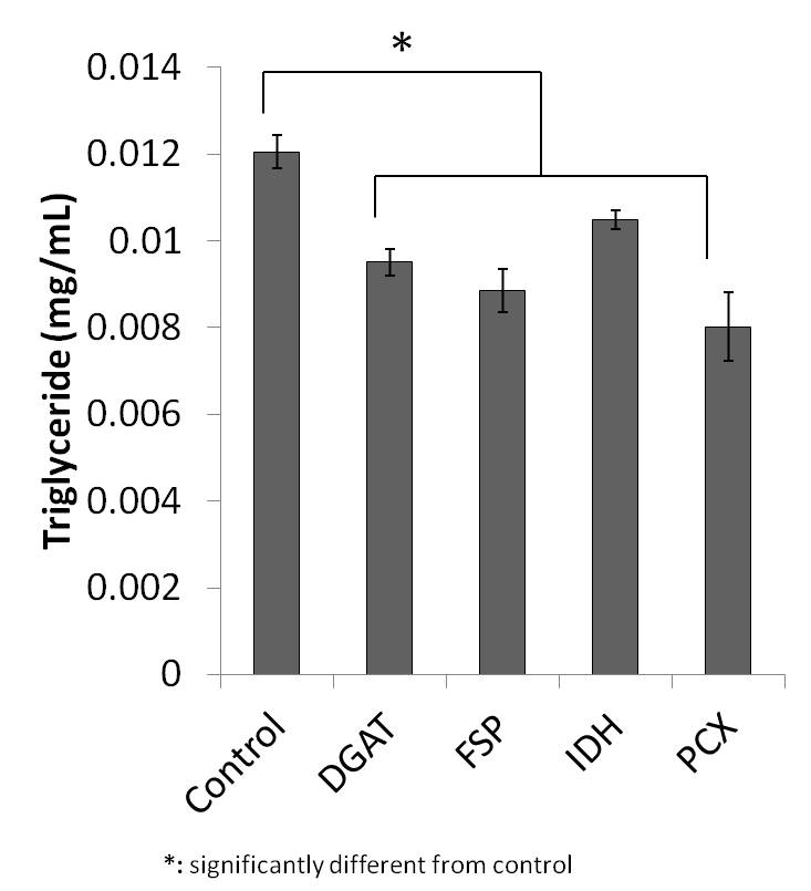
Fig 1 Identification of knockdown targets in 2D planar monoculture.
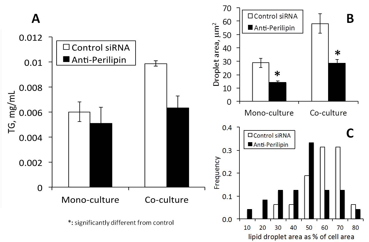
Fig 2 (A) Triglyceride accumulation in 3D mono- and coculture treated with non-targeting (control) or anti-perilipin siRNA. Error bars are SEM. (B) Average lipid droplet size in 3D mono- and coculture treated with non-targeting (control) or anti-perilipin siRNA. Error bars are SEM. *Significantly different from respective control (n~1,000 droplets from 8 different images), p=0.0005 and p=0.03, respectively. (C) Histogram of droplet size in 3D monoculture.
See more of this Group/Topical: Food, Pharmaceutical & Bioengineering Division