
 Google
 Yelp
 Facebook
 Twitter
 ChEnected
342194 Characterization of the Permeability of the Brain Endothelium Due to Electroporation Using a Dynamic Microengineered Model
Abstract
The microvascular endothelial cells, which cover the interior of the brain microvascular network, play a significant role in controlling the transport of different molecules into the central nervous system (CNS). Although crucial to human health by maintaining the neuroparenchymal microenvironment and protecting the neural tissue from toxins, the blood-brain barrier (BBB) also limits the success rate of new therapies. One significant obstacle to drug development for targeting the CNS has been the high resistivity of the BBB in allowing large molecules to penetrate into the CNS. Electroporation, which is the permeabilization of the cell membrane using pulsed electric fields, is a non-viral physical method for drug delivery [1]. We fabricated a microengineered BBB model and evaluated its permeability to different chemicals via delivery of pulsed electric fields by using a novel laminated microdevice with embedded electrodes which accommodates the BBB mimic. Our results support the hypothesis that electroporation is able to increase the permeability of the BBB without compromising cell viability in an in vitro model.
Introduction
Organs on a chip, which are a class of microdevices that feature a physiologically relevant function, are a method of in vitro tissue analysis. The BBB has a significant role in screening certain molecules from the CNS and is a major obstacle to drug development for many CNS diseases. Hence, many researches are focused on studying the physiology of this barrier and how to permeate through it. Booth et al. [2] developed a microfluidic BBB model incorporating a co-culture of endothelial and astrocytes and monitored the formation of tight junctions using transendothelial electrical resistance. In another study Prabhakarpandian et al. [3] modeled a micro BBB using brain endothelial cells in astrocyte conditioned medium and investigated the effect of astrocytes on the formation of the tight junctions and the transport of certain chemicals across the barrier. Flow induced shear force and chemical factors are also deployed to modulate the permeability of the BBB [4]. In this study, we stimulated the microengineered BBB model by short pulsed electric fields (BTX ECM 830, Harvard Apparatus)) to induced cell electroporation and disrupt the blood-brain barrier. The transport of chemicals across the BBB was quantified as a function of molecular weight, electric field and flow rate.
Materials and Methods
The microfluidic device was fabricated using photolithography and replication molding. A polycarbonate membrane was embedded into the device separating an upper and lower chamber (100µm deep and 300 µm wide). Rat brain endothelial cells (RBE4) were cultured on the membrane until the formation of a confluent monolayer and the expression of the tight junctions. The microdevice features a pair of microelectrodes on one side of the barrier. By applying electric pulses of different amplitudes, the endothelial cells were reversibly electroporated and the permeability of the barrier was changed. We injected Dextran (Sigma) of different molecular weights into one chamber and measured the diffused amount into the other chamber following electroporation treatments.
Results and Discussion
Brain endothelial cells were cultured on the porous membrane inside the microenvironment until a confluent monolayer of cells was formed. We repeated the experiment with the presence of astrocytes on the opposite side of the membrane. Astrocytes are star shaped glial cells in the brain and spinal cord which support the endothelial cells and contribute to the formation of the tight junctions. To have a better representation of the in vivo conditions, media was perfused over the endothelial cells in the top chamber. Figure 1 shows a schematic of the experimental setup. By applying electric pulses between the electrodes, the permeability of the barrier was temporarily increased, allowing certain molecules of high molecular weight to penetrate through the BBB. The permeability analysis after the electroporation and live/dead assay confirms the reversibility of the process without compromising cell viability.
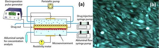
Figure 1. (a) Test setup including the BBB mimic, electrical, and flow circuits. Two halves of the microfluidics device fabricated in glass and PDMS are bonded together, encapsulating the membrane and forming separate luminal and abluminal channels in both sides of the membrane. (b) a monolayer of RBE4 cells cultured in the microchannel of the device.
Conclusions:
We modeled the BBB inside the microfluidic device and stimulated it by electroporation pulses to increase its permeability to certain chemicals without affecting cell viability. Using this method we are able to quantify the required electric field for delivery of certain drugs in a dynamic environment.
References:
[1] Garcia, P. et al, PLoS ONE, 2012, 7(11), e50482
[2] Booth, R. et al, Lab Chip, 2012, 12, 1784.
[3] Prabhak, B. et al, Lab Chip, 2013, 13, 1093.
[4] Griep, L. Biomed. Microdevices, 2013, 15, 145-150.
See more of this Group/Topical: 2013 Annual Meeting of the American Electrophoresis Society (AES)