
 Google
 Yelp
 Facebook
 Twitter
 ChEnected
342521 Zyxin Morphology, Concentration and Arrangement Modulates Force Magnitude and Directionality in Nanofibrous Environment
Zyxin Morphology, Concentration and Arrangement Modulates Force Magnitude and Directionality in Nanofibrous Environment
Colin Ng and Amrinder S. Nain
Department of Mechanical Engineering, Virginia Tech, Blacksburg, VA
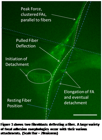
| Figure SEQ Figure \* ARABIC 1 shows two fibroblasts deflecting a fiber. A large variety of focal adhesion morphologies occur with their various attachments. (Scale Bar = 20micorns) |
 Introduction: In the human body, cells attach
to and receive biophysical cues from the surrounding fibrous microenvironment
known as the extracellular matrix (ECM). These biophysical cues play an active
role in cell morphogenesis, migration, adhesion, proliferation, differentiation
and complex inflammatory cascades. However, the exact mechanism by which the cell
interacts and responds to the ECM mechanistic environment is not well
understood. Recent cellular force measurement
techniques including pillars, cantilevers, gels etc., have shown unique focal
adhesion morphologies with different force generating behaviors. In this study,
we further this understanding by demonstrating the role of zyxin
arrangement in force modulation in a suspended nanofibrous environment.
Materials and Methods: Using STEP (Spinneret based Tunable Engineered Parameters) fiber manufacturing platform, we are capable of measuring the forces of migratory cells along suspended fibers ranging from sub 100nm – micron in diameter. Using an in-house solvent setup, multiple layer fibers are fused together at intersecting points, thus, creating a customized nanonet of fiber networks. In this system, the forces are easily measured by recording the deflection of fiber and using Euler beam mechanics in the elastic limit. Focal adhesion structure and arrangement are studied through time-lapse imaging of NIH3T3 fibroblasts tagged with zyxin and its relation to force.
Results: As shown in the Figure 1, a variety of unique focal adhesion arrangements are observed during force modulation of NIH3T3 fibroblast. Maximum force is exerted when the zyxin proteins align themselves with the deflecting fiber into highly concentrated clusters. Detachment is initiated when the adhesions disperse and unaligned themselves with the fiber. This continues until only a few elongated adhesions remain and eventually completely detach from the fiber.
Conclusion: Zyxin morphology, concentration and arrangement fuctions in determining force directionality and magnitude in a migratory cell in a suspended fibrous environment. Utilizing this knowledge of zyxin's special and temporal role in force modulation can be used to develop diagnostic and drug testing platforms.
See more of this Group/Topical: Food, Pharmaceutical & Bioengineering Division