
 Google
 Yelp
 Facebook
 Twitter
 ChEnected
342582 A Multiscale Approach to Stem Cell-Based Chondrogenesis for Cartilage Repair
Tissue engineering is a promising method for long-term repair of cartilage lesions; however, current tissue-engineered cartilage constructs have inferior mechanical properties compared to native cartilage. Other researchers attribute this problem to a lack of an oriented structure in the constructs at the microscale that is present in the native tissue. In this study, we utilized contact guidance to develop constructs with microscale architecture for improved chondrogenesis and function. In this system, we used microscale channels on the scaffold surface to guide mesenchymal stem cells (MSCs) undergoing chondrogenesis to produce oriented extracellular matrix (ECM). Stable channels of varying microscale dimensions were formed in collagen-based and polydimethylsiloxane membranes via a combination of microfabrication and soft-lithography. Human MSCs were selectively seeded in these channels. The chondrogenic potential of MSCs seeded in these channels was investigated by culturing them for 3 weeks under differentiating conditions with transforming growth factor β1 and then evaluating the subsequent synthesized tissue for mechanical function and by type II collagen immunohistochemistry. We demonstrated selective seeding of viable MSCs within the channels (Fig. 1). MSC aligned and produced mature collagen fibrils along the length of the channel in smaller linear channels of widths 25-100 µm compared to larger linear channels of widths 500-1000 µm. Further, substrates with microchannels that led to cell alignment also led to superior mechanical properties compared to constructs with randomly seeded cells or selectively seeded cells in larger channels. The ultimate stress and modulus of elasticity of constructs with cells seeded in smaller channels increased by as much as four folds (Fig. 2). We demonstrated that microscale guidance is useful to produce 2-dimensional oriented cartilage structures with improved mechanical properties. Furthermore, we extended the 2-dimensional construct findings to a 3-dimensional construct for preclinical experiments. We successfully created 3-dimensional constructs for in vivo applications by rolling up 2-dimensional MSC-seeded collagen based scaffolds and formed 3-dimensional rolled-up large scale (3.5 mm in diameter × 18 mm in length) cartilage constructs with microscale architecture to guide the differentiating MSCs to produce oriented ECM. Histology and immunohistochemistry indicated extensive glycosaminoglycan and collagen type II production in the 3-dimensional constructs, which are both indicative of chondrogenesis (Fig. 3). Our results show that the microscale guidance channels incorporated within the 3-dimensional cartilage constructs led to the production of aligned cell-produced collagenous matrix and enhanced mechanical function. The tissue modulus of elasticity of the 3-dimensional cartilage constructs containing guidance channels (1.16-2.07 MPa) increased by as much as six times compared to constructs without channels, which are higher than most current tissue-engineered constructs (Fig. 4). Overall, these findings offer new insight into how microscale guidance channels can regulate matrix deposition and long term construct development. Furthermore, our results provide new parameters which help optimize the design of functional MSC-based tissue-engineered cartilage using collagen-based substrates and can be used to fabricate large clinically useful MSC-based cartilage constructs with superior mechanical properties.
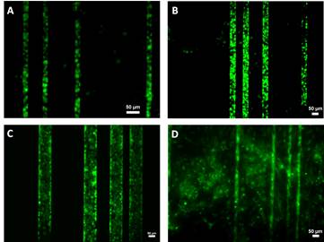
Fig. 1 Fluorescent images of cells in linear channels of widths 25 μm (A), 50 μm (B), and 100 μm (C) show selective seeding compared to untreated channel surfaces (D). Scale bars: 50 μm
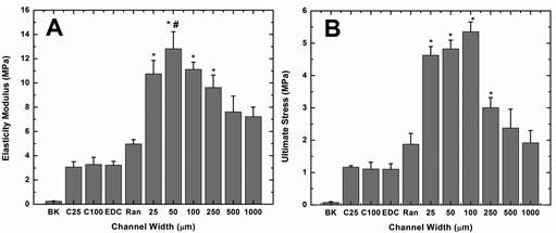
Fig. 2 Effect of microscale guidance on mechanical properties. Collagen membranes were subject to tensile testing until failure. Mean values of elasticity modulus (A), and ultimate stress (B) are shown. BK and EDC represent respectively uncrosslinked and EDC crosslinked collagen membranes without microchannels or MSCs. C25 and C100 represent collagen membranes consisting of 25 μm and 100 μm linear channels only, without MSCs. All other channels were seeded with MSCs. Ran: EDC crosslinked collagen membranes without channels; 25, 50, 100, 250, 500 and 1000 represent widths (μm) of linear channels in the EDC crosslinked collagen membranes (A) * indicates statistically significant difference (p < 0.05) compared to data with no channels (RAN). # indicates statistically significant difference (p < 0.05) compared to data with 1000 μm channels (1000). (B) * indicates statistically significant difference (p < 0.05) compared to data with no channels (RAN), 250 (250), 500 (500) and 1000 μm (1000) -wide channels.
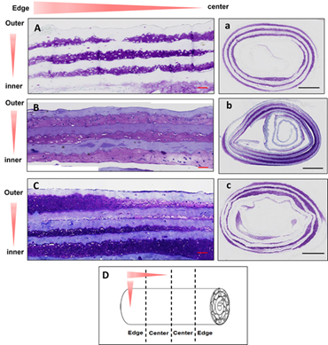
Fig. 3 Cartilage tissue constructs formed by the role up process. Three different microchannel designs were used. A-C indicate longitudinal sections and a-c indicate cross sections. Scale Bar=100 μm (A-C), 1 mm (a-c)
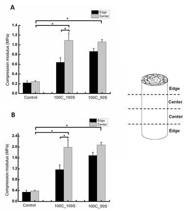
Fig. 4 Mechanical properties of cartilage constructs with microchannel guided ECM. Compressive modulus of the whole construct (A) and the chondrogenic tissue (B) are shown.
See more of this Group/Topical: Food, Pharmaceutical & Bioengineering Division