
 Google
 Yelp
 Facebook
 Twitter
 ChEnected
342657 Single Nanofiber Structural Stiffness Directly Affects Cellular Migration and Cytoskeletal Response of C2C12 Cells
Introduction
Cell substrate interactions are an important part of many biological processes, including wound healing, stem cell differentiation, and immune response [1,2]. Both the cytoskeletal arrangement and migration characteristics respond to physical and chemical cues from the cell substrate in each of these processes. The substrate that cells are interacting with while in vivo is the extracellular matrix (ECM), a dense fibrous mesh that serves as the support structure for tissues [3–5]. Previous in vitro studies have adopted the use of both fibrous constructs and gels made of collagen, fibrin, or other biocompatible materials in order to try to match the substrate interactions that are seen between the cell and ECM. However, there has been little research into the effects of the local stiffness of the microarchitecture that makes up these fibrous substrates on cell behavior, despite the fact that the overall material modulus of the substrate has been shown to alter cellular behaviors, including cytoskeletal arrangement and migration characteristics [6–8]. In this study the previously reported STEP technique (Spinneret based Tunable Engineering Parameters) has been utilized to create evenly spaced single suspended nanofibers on which the effects of the gradient of fiber structural stiffness has been explored [9,10]. These studies in understanding single cell mechanics will aid in design of nanostructured biomaterials.
Methods and Materials
Aligned, evenly spaced single polystyrene nanofibers were deposited on a substrate consisting of two polydimethysiloxane (PDMS) blocks fixed to a glass slide. Before fiber deposition, PDMS constructs were coated with uncured PDMS to act as an adhesive. Fibers were coated with 2 µg/ml fibronectin overnight before C2C12 mouse myoblasts were cultured on the fibers. Cells adhered to single nanofibers and migrated along the fiber length. Time lapse imaging was performed to allow for migration speed and cell spreading characterization. Cells were fixed using 4% paraformaldehyde and immunostained for focal adhesions, f-Actin stress fibers, and the nucleus. Characterization of cell length, focal adhesion cluster length, and nuclear shape index was performed using immunostained images.
Results and Discussion
It has been found that cells sense and respond to the changing gradient of structural stiffness that is present along a single nanofiber. Cell length and focal adhesion cluster length have been shown to increase with increasing structural stiffness, while nuclear shape index has been shown to decrease with increasing structural stiffness [11] Additionally, preliminary results have shown that cells reach their fully spread state and begin migrating in less time on areas of higher structural stiffness. These modifications to the cytoskeletal arrangement and cellular behavior in turn lead to a decrease in average cellular migration speed at higher structural stiffness areas as shown in Figure 1.
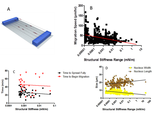
Figure 1. A) Typical suspended nanofiber substrate consisting of a glass coverslip with fibers deposited over and fixed to two PDMS blocks. B) Migration speed reaction to structural stiffness. Average migration speed decreases with increasing structural stiffness. C) Time for cells to reach their fully spread state and begin migrating decreases with increasing structural stiffness. D) Nucleus length and width respectively increase and decrease with increasing structural stiffness.
Conclusions
The results found in this work indicate that cells are employing a substrate stiffness sensing mechanism that goes beyond bulk material stiffness sensing. Cells are shown to respond to structural stiffness by modulating their cytoskeletal characteristics both in magnitude and temporally leading a change in migratory behavior. These results add to the current understanding of single cell mechanics and aid in design of nanostructured biomaterials.
References
[1] Evans N. D., Minelli C., Gentleman E., LaPointe V., Patankar S. N., Kallivretaki M., Chen X., Roberts C. J., and Stevens M. M., 2009, “Substrate stiffness affects early differentiation events in embryonic stem cells.,” European cells & materials, 18, pp. 1–13; discussion 13–4.
[2] Wang Y., Wang G., Luo X., Qiu J., and Tang C., 2012, “Substrate stiffness regulates the proliferation, migration, and differentiation of epidermal cells.,” Burns : journal of the International Society for Burn Injuries, 38(3), pp. 414–20.
[3] Lo S. H., 2006, “Focal adhesions: what's new inside.,” Developmental biology, 294(2), pp. 280–91.
[4] Reilly G. C., and Engler A. J., 2010, “Intrinsic extracellular matrix properties regulate stem cell differentiation.,” Journal of biomechanics, 43(1), pp. 55–62.
[5] Frantz C., Stewart K. M., and Weaver V. M., 2010, “The extracellular matrix at a glance.,” Journal of cell science, 123(Pt 24), pp. 4195–200.
[6] Mitrossilis D., Fouchard J., Pereira D., Postic F., Richert A., Saint-Jean M., and Asnacios A., 2010, “Real-time single-cell response to stiffness.,” Proceedings of the National Academy of Sciences of the United States of America, 107(38), pp. 16518–23.
[7] Ghosh K., Pan Z., Guan E., Ge S., Liu Y., Nakamura T., Ren X.-D., Rafailovich M., and Clark R. a F., 2007, “Cell adaptation to a physiologically relevant ECM mimic with different viscoelastic properties.,” Biomaterials, 28(4), pp. 671–9.
[8] Schoen I., Hu W., Klotzsch E., and Vogel V., 2010, “Probing cellular traction forces by micropillar arrays: contribution of substrate warping to pillar deflection.,” Nano letters, 10(5), pp. 1823–30.
[9] Nain A. S., Sitti M., Jacobson A., Kowalewski T., and Amon C., 2009, “Dry Spinning Based Spinneret Based Tunable Engineered Parameters (STEP) Technique for Controlled and Aligned Deposition of Polymeric Nanofibers.,” Macromolecular rapid communications, 30(16), pp. 1406–12.
[10] Nain A. S., Phillippi J. a, Sitti M., Mackrell J., Campbell P. G., and Amon C., 2008, “Control of cell behavior by aligned micro/nanofibrous biomaterial scaffolds fabricated by spinneret-based tunable engineered parameters (STEP) technique.,” Small (Weinheim an der Bergstrasse, Germany), 4(8), pp. 1153–9.
[11] Meehan S., Nain A.S., 2013, "Effect of Nanofiber Stiffness on C2C12 Myoblast Migration." In Review
See more of this Group/Topical: Materials Engineering and Sciences Division