
 Google
 Yelp
 Facebook
 Twitter
 ChEnected
342707 Nanofiber Based Cellular Protrusions of Cancerous Cells
Nanofiber based Cellular Protrusions of Cancerous Cells
Brian Koons1, Puja Sharma2, Amrinder S. Nain1,2
1Mechanical Engineering Department, Virginia Tech, Blacksburg, USA
2School of Biomedical Engineering and Sciences, Virginia Tech, Blacksburg, USA
Introduction
As cells migrate and interact with their mechanical environment of the extracellular matrix (ECM), they sense their surroundings using cytoplasmic protrusions. Generally, these protrusions can be classified as either lamellipodia or filopodia, where actin within the protrusion is in the form of a broad branched sheet or thin long bundle, respectively [1]. Although lamellipodia have been known to play a pivotal role on 2D flat substrates, filopodia proves to be the dominant sensing mechanism in 3D microenvironments [2]. In cancerous cells, filopodia-like projections known as invadopodia are used to break down the surrounding ECM and infiltrate the proximate blood vessels. Current studies consist of using chemical gradients to force cancer cell protrusions across a porous membrane. Here, alternatively, we demonstrate a biophysical method using nanofibers to selectively position areas of contrasting structural stiffness (N/m) in order to elicit cellular protrusions. Using the previously reported STEP technique, non-electrospun suspended polymer nanofibers are deposited on substrates with varying fiber diameters in aligned configurations to create stiffness and geometric gradients sensed by the cell [3]. Cells interacting with this system are seen to migrate on the stiff fibers while sending protrusions towards and on the less stiff environment of nanofibers. Therefore, this method when coupled with immunostaining, can provide an understanding of the surrounding biophysical influence on actin polymerization and depolymerization. Use of this method provides a protrusion-based comparison for the invasiveness and metastatic potential of various cancer cell types including brain, breast, and prostate.
Materials and Methods
Nanofibers were fabricated from polymer solutions of polystyrene dissolved in xylene using various concentrations and molecular weights. The fibers were suspended in a highly controlled and aligned fashion across polystyrene frames via STEP technique to create a fibrous grid where crossing fibers are perpendicular to one another. Vertical fibers averaged 2 µm in diameter while horizontal fibers were 250 nm or 500 nm in diameter for each sample. Diameter measurements were confirmed using scanning electron microscopy. Fusing fibers at their intersections created unified fiber meshes with both fibers coexisting on the same geometric plane. The fiber meshes were coated with ECM protein fibronectin (2 µg/ml) to promote cellular adhesion prior to cell seeding with Denver Based Tumor Research Group (DBTRG-05MG) glioma cell line. Time-lapse imaging and measurements were performed using Carl Zeiss microscope and Axiovision software respectively. As cells initiated protrusions throughout the nanofiber mesh, protrusion length (µm), extension rate (µm/min), and retraction rate (µm/min) were measured using aforementioned software.
Results
Protrusion Behavior
DBTRG-05MG favors the 2 𝜇m fibers, and mostly stayed wrapped around the 2 𝜇m fibers due to high structural stiffness. Cells rarely migrated on less stiff nanoscale fibers. Instead, the cells extended protrusions from the stiff microfibers onto the less stiff, smaller diameter nanofibers, as shown in Figure 1. These protrusions were observed to be thin rod-like cytoplasmic projections, and their occurrence was observed to depend on the fiber diameter upon which they extended. Cells were observed to extend multiple protrusions in opposing directions, sensing potential migratory pathways within distance of contact.
Protrusion Analysis
The average protrusion length of 23 µm was constant between both 250 nm and 500 nm fibers. In addition, the protrusion extension rate of 3 µm/min did not differ between diameters. However, the rate of protrusion retraction for the larger diameter fibers (3.3 µm/min) was slower than when compared to retraction rates on smaller diameter fibers (4.2 µm/min) as shown in Figure 1.
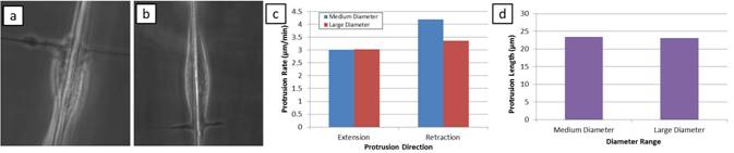
Figure 1. (a) Cell protrusion on large, 500 nm fiber, (b) bi-directional cell protrusion on medium, 250 nm fiber, (c) protrusion extension and retraction rates between large and medium fiber diameters, and (d) total length of protrusion compared between large and medium fibers.
Discussion
The study shows that biophysical characteristics of the extracellular microenvironment can control cell attachment and migration preference. Preliminary data illustrates that retraction can be regulated through the variance of fiber diameter. However, protrusion length and extension rates were seen to be constant on the diameters tested. Although the protrusions are sensing the mechanical characteristics of the extracellular environment, they can also be seen as migratory attempts to overcome the prohibitory stiffness gradient which is keeping the cell migrating along the large, stiffer fiber. Their protrusion extending potential can quantify the invasiveness and metastatic potential of cancerous cells. Our future experiments are aimed at exploring protrusion dynamics for a variety of cancer and normal cell lines on fibers of different diameters and structural stiffness.
References
[1] Faix, Jan Breitsprecher, Dennis Stradal, Theresia E B Rottner, Klemens. Filopodia: Complex models for simple rods. The international journal of biochemistry & cell biology. 2009
[2] Albuschies, Jörg Vogel, Viola. The role of filopodia in the recognition of nanotopographies. Scientific Reports. 2013
[3] Nain AS, Sitti M, Jacobson A, Kowalewski T, Amon C. Dry Spinning Based Spinneret Based Tunable Engineered Parameters (STEP) Technique for Controlled and Aligned Deposition of Polymeric Nanofibers. Macromolecular Rapid Communications. 2009;30:1406-12.
See more of this Group/Topical: Food, Pharmaceutical & Bioengineering Division