
 Google
 Yelp
 Facebook
 Twitter
 ChEnected
342827 Modular Biomaterial Scaffolds for Scalable Tissue Assembly and Rapid Vascularization
Statement of Purpose: Tissue engineering aims to create functional biological tissues to treat diseases and damaged organs. A primary goal is to fabricate 3D constructs that can promote cell-cell interactions and tissue level organization. Accomplishing these prerequisites with the currently available conventional scaffolds and fabrication techniques still remains a challenge. To reproduce the full functionality there is a need to engineer tissue constructs that mimic the innate architecture and complexity of natural tissues. The emerging field of modular tissue engineering aims to fabricate more efficient and complex tissue constructs from the bottom up, with the desire to recreate the native architecture and to promote extensive vasculature.
Using this strategy, we propose to develop methods for fabricating modular tissue constructs by assembling ECM based microscale modules. These biodegradable modules possess tunable interior environments, can be seeded internally and externally, and can be assembled into tissue constructs with parenchymal and vascular components. The proposed work is based on the hypotheses that the modular constructs assembled from micro scale modules permeated by a network of interconnected, endothelial cell-lined channels can facilitate extensive vascularization and mass transport. This work sought to develop methods for assembling 3D constructs by fusing microcapsules reloaded with biopolymer solution with the long term goal of engineering functional tissues by establishing a technology foundation for subsequent rapid assembly of three-dimensional, tissue density constructs.
Methods: Porcine aortic smooth muscle cells (SMC) and Rat hepatocytes were encapsulated in microcapsules produced by complex coacervation between chitosan, collagen and hyaluronan. Briefly, cells were suspended in a polyanion solution (0.5 wt% hyaluronan, 0.1 wt% type I collagen, 3.8 wt% sorbitol). Droplets of this solution were generated and collected in stirred chitosan solution (0.6 wt% high MW chitosan, 3.8 wt% sorbitol, 1 wt% acetic acid). Ionic interactions between oppositely charged polymers at the droplet surface generated the encapsulating membrane. To explore culture of fused capsule modules, 1 week cultured SMC capsules were equilibrated with 1 wt% heparin in PBS for 2 hours. They were then fused by briefly rinsing with 0.06 wt. % chitosan solution and allowing 4-5 capsule layers to sit in contact for 10 minutes, followed by culture medium equilibration. The capsule with hepatocytes in addition was spun for 10 seconds in 50G, to take out the excess solution. Separate cultures explored the external seeding of porcine aortic endothelial cells (EC) on outer surfaces.
Results/Discussion: Hyaluronan-collagen containing capsules exhibited cell-mediated contraction during culture, resulting in the elimination of internal void space within capsules. H&E staining of sections from the fused, multilayered capsule constructs revealed a compact internal structure with inter-capsule channels capable of supporting promote vasculogenesis (Fig. A). Trichrome staining of these sections showed a dense collagen matrix inside capsules and also large amount of collagen integrated into the capsule walls (Fig. B). This irregular collagen rich outside wall may provide a superior substrate for support of extracapsular angiogenesis. The sections of the hepatocyte seeded construct showed more densely packed cell construct with potential vascularizable spaces. The EC seeded onto SMC containing capsules exhibited extensive proliferation (Fig. D). SMC in these co-culture capsules also exhibited higher rates of proliferation compared to SMC-only capsules. Hence, this approach has the potential to make vascularized tissue constructs with high tissue densities.
A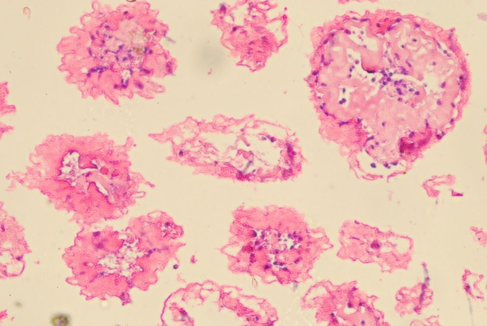 B
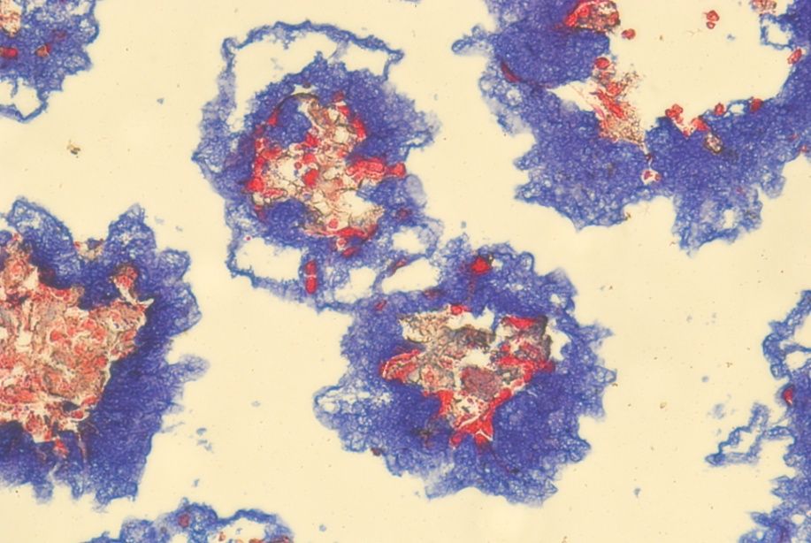
C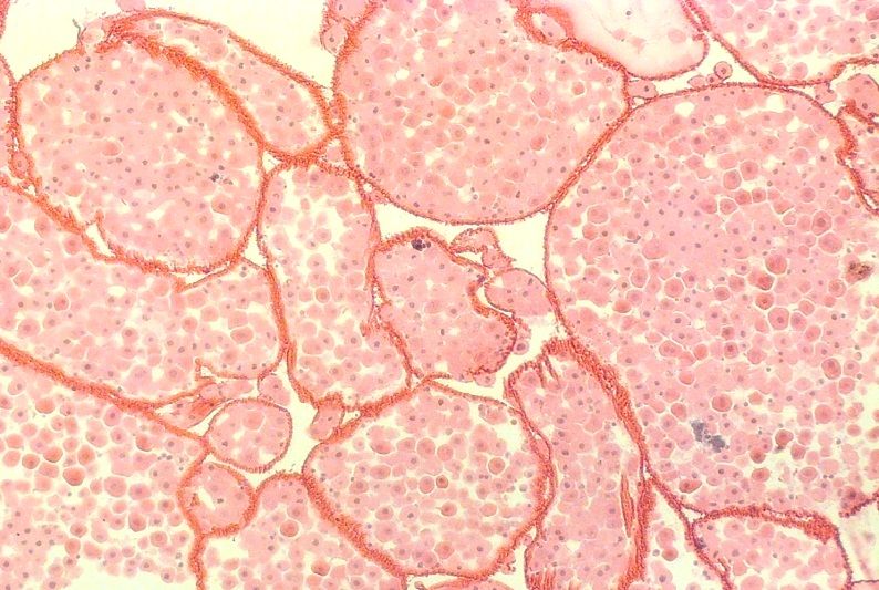 D
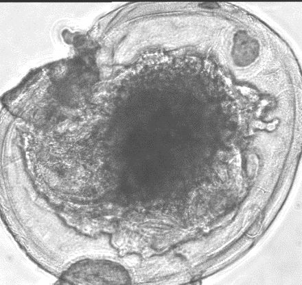
Conclusions: These results demonstrate that GAG-based microcapsules can be fused to from 3D constructs with vascularizable interconnected channels. This finding establishes a technology foundation for subsequent rapid assembly of three-dimensional, tissue density constructs. When coupled with growth of endothelial cells on the external capsule surfaces, these scalable systems are a promising platform for modular tissue engineering of several organ systems.
See more of this Group/Topical: Materials Engineering and Sciences Division