
 Google
 Yelp
 Facebook
 Twitter
 ChEnected
343430 Aortic Smooth Muscle Cell Benchmark Force Measurement
Aortic Smooth Muscle Cell Benchmark Force Measurement
Brian Koons1
Julie A. Phillippi2
Thomas G. Gleason2
Amrinder S. Nain1,3
1Department of Mechanical Engineering, Virginia Tech, Blacksburg, VA, USA
2 Department of Cardiothoracic Surgery, University of Pittsburgh, Pittsburgh, PA, USA
3School of Biomedical Engineering and Sciences, Virginia Tech, Blacksburg, VA, USA
Introduction
Rupture of aortic aneurysm is the thirteenth leading cause of death in the United States and is a clinical emergency. The main reason for the high death rate is that the condition is commonly asymptomatic, until the aorta ruptures, leading rapidly to death. An aortic aneurysm is characterized by an enlargement of the aorta's diameter. Although vessel diameter varies among individuals, it can be related to body surface area, and aortic diameter larger than 30 mm is concerning and potentially life threatening [1]. Aortic aneurysm can be either fusiform, affecting the entire circumference of the vessel, or saccular, where only a portion of the circumference is enlarged. The dilatation is due to mechanical weakening of the blood vessel wall, initiated by biological pathways that are distinct for aneurysms of the thoracic and abdominal aorta. Current expert clinical opinion dictates surgical intervention to replace the aneurysmal aorta with a synthetic graft when the maximal orthogonal aortic diameter reaches 50-55 mm. Unfortunately, the aneurysmal aorta has been known to dissect or rupture at lower aortic diameters and accurate risk assessments for patients is challenging and lacking. Hence, there is a great clinical need for enabling technologies to improve rupture risk profiles, increased knowledge of the biological mechanisms mediating the pathophysiology and new medical therapies for patients affected by aneurysm. We have engineered a model system used to measure forces exerted by migratory primary human aortic smooth muscle cells (HASMC). This technology could be used as a benchmark to determine putative differences in cell forces exerted by diseased HASMCs. Using our previously reported STEP (Spinneret based Tunable Engineered Parameters) non-electrospinning fiber fabrication platform [2], aligned polymeric fibers were deposited in multiple perpendicular layers and forces exerted by migratory cells were calculated through the deflection of fibers in the elastic limit using Euler beam mechanics. This platform will provide fresh insights in single-cell force generation in normal vs. diseased cells and can potentially be used to determine efficacy of novel therapeutics to treat patients at risk for aortic catastrophe.
Materials and Methods
STEP manufactured nanofibers were suspended in a highly aligned and oriented fashion on polystyrene frames. Various concentrations of polystyrene dissolved in xylene was used to fabricate fibers of 250 nm and 2 𝜇m, respectively. The two fiber types were oriented perpendicular to one another and fused at intersections to engineer a nanofiber mesh where both fiber diameters coexist within the same 2-D geometric plane. The fibers were coated with the extracellular matrix protein fibronectin (2 𝜇g/ml) to increase cell attachment (seeding density 20,000 cells/ml). HASMCs were isolated from non-aneurysmal patients with approval of the IRB and with informed patient consent. Time-lapse phase contrast microscopy and forces measurements were conducted using Carl Ziess microscope and Axiovision software. As cells migrated on the fibers, fiber deflections were observed and measured to calculate the cell forces using Euler beam theory.
Results
Cellular force generation was found to be dependent upon fiber structural stiffness (N/m). The cells were unable to deflect the stiffer 2 𝜇m diameter fibers. However, large deflections, ranging from 2 to 12 𝜇m, occurred on the less stiff 250 nm fibers. Therefore, the less stiff fibers can be modeled as deformable beams where the stiff fibers at their edge create a simply supported boundary condition. Preliminary results with HASMC exhibited a force generation of 0.01 nN at the least stiff regions along the fiber and up to 0.8 nN near the stiffest regions of the fiber, as shown in Figure 1. These measurements provide the benchmark values for non-diseased HASMC lines and can be used to compare force generation by cells from diseased patients.
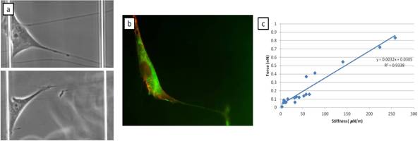
Figure 1. (a) Healthy HASMC deflecting 250 nm fiber segments between stiff boundaries, (b) immunocytochemistry for the cytoskeletal proteins F-actin (red) and zyxin (green) while a healthy HASMC is deflecting a fiber, and (c) healthy HASMC force generation on fibers of various stiffness.
Discussion
The STEP fiber fabrication technique provides a novel biophysical platform for determining the force generation of single migratory HASMCs. The method may be used to establish a new force-based hypothesis for aneurysm development, progression and risk for rupture and will help test the efficacy of novel therapeutics. It is expected that future studies on diseased cells will show a decrease in cell force generation for HASMC isolated from aneurysmal patients when compared with healthy HASMC. This enabling technology can later be applied for the study of several other pathologies.
References
[1] Sakalihasan, N Limet, R Defawe, O D. Abdominal aortic aneurysm. Lancet. 2005
[2] Nain AS, Sitti M, Jacobson A, Kowalewski T, Amon C. Dry Spinning Based Spinneret Based Tunable Engineered Parameters (STEP) Technique for Controlled and Aligned Deposition of Polymeric Nanofibers. Macromolecular Rapid Communications. 2009;30:1406-12.
See more of this Group/Topical: Nanoscale Science and Engineering Forum