
 Google
 Yelp
 Facebook
 Twitter
 ChEnected
343822 Simulations of CSF Flow Dynamics in a Global CNS Model With Magnetically Targeted Intrathecal Drug Delivery
Introduction: Therapeutic intervention for central nervous system diseases has limited effectiveness due to drug transport restriction across the blood brain barrier. Intrathecal (IT) drug delivery aims to circumvent this obstruction, however interpatient variability impacts treatment outcomes. Drug distribution is affected by patient anatomy, cerebrospinal fluid (CSF) properties (volume, pressure, and density), heart rate, and drug infusion properties. A secondary problem with IT drug delivery is localization of the compound to a region of interest. External non-invasive magnetic fields have been shown to direct and concentrate magnetic nanoparticles for treatment of several diseases such as tumors. We propose to understand and explore the efficacy of treating CNS diseases with IT administration of magnetic nanocarriers functionalized with therapeutic molecules. Multi-phase simulations of a complete patient specific model of the CSF filled spaces can predict dispersion patterns of nanoparticle-therapeutic complexes both in a native state and under magnetic field influence. An in vitro bench top human spinal canal model with magnetic field guidance is used to corroborate computational simulation results.
Computational CSF Fluid Flow Methods: Phase contrast magnetic resonance imaging (PC-MRI) in combination with computational fluid mechanics is beginning to provide a complete picture of patient specific CSF flow fields. Patient specific MRI data allows for generation of an anatomically consistent mesh for computer modeling giving accurate predictions of drug dispersion patterns in the CNS.Modeling of spinal CSF flow is focusing on the role of the micro-anatomy including nerve roots and trabeculae. Using simulations, our lab showed that CSF pulsatility and nerve roots play a significant role in the bio-distribution of drug molecules during intrathecal drug delivery. Our work also illustrates the formation of complex micro-mixing patterns which are attributable to nerve roots and trabeculae.
We constructed a patient-specific model of the fluid filled spaces inside the CNS based on PC-MRI data. Pulsatile motion of the CSF for the full model is generated by explicitly moving the choroid plexus and parenchyma according to a Fourier series that captures the velocity wave of the basilar artery. The model includes regions of production and absorption at the choroid plexus and arachnoid villi respectively. Nerve roots were reconstructed from the image data and incorporated into the full SAS model. We added micro-anatomical detail below the image resolution threshold of MRI to quantify the effect of trabeculae (6 micron diameter) on CSF flow. We compared complex flow profiles to simulations in structures lacking the anatomical detail. The effect on spinal flow resistance and their impact on complex flow profiles as a function of varying trabeculae density. The full spinal SAS model visualizes flow profiles at 31 nerve pairs over a complete cardiac cycle predicting the effect of successive nerve roots.The computational fluid dynamic solver ANSYS Fluent 14.0 is used for multi-phase fluid flow simulations to predict dispersion of magnetic nanoparticle based drug infusate within the spinal canal. Magnetic fields of varying strength were also applied to the complete CNS model simulations to understand the effect on magnetic nanocarrier transport and accumulation in a global model.
Bench top Testing Methods of Magnetic Nanoparticle Guidance: Most intrathecally injected drugs are required only at certain locations within the spinal canal where it is essential for these drugs to diffuse into the spinal cord to produce a therapeutic effect. Currently, in order to reach the crucial drug concentration at the target site and aid diffusion into the spinal cord, high drug doses are used. With no method to deliver the large drug doses locally, current intrathecal drug delivery treatments are hindered with wide drug distributions throughout the CNS which cause harmful side effects. Our lab proposed a magnetic nanoparticle based drug delivery technique within the CNS by IT administration. An externally applied magnetic field localizes the magnetic drug coated nanoparticles at desired locations. Detailed simulations in COMSOL Multi-physics optimized the placement and strength of the external magnet. A physiologically consistent bench top model of the human spinal canal was constructed to determine the effect of magnetic fields on magnetic nanoparticle distribution, shown in Figure 1.
|
Figure 1: Schematic of entire experimental setup with the in vitro human spine model which clearly shows the three different zones (injection, targeting, and barrier zones). |

A co-precipitation technique using ferrous and ferric salts was used to form the 8-10 nm diameter superparamagnetic magnetite nanoparticle cores, which were then coated with a highly functionalizable gold layer of thickness between 8-15 nm. The nanoparticles were characterized by TEM, EDS, x-ray diffraction spectroscopy and SQUID magnetometry. The in vitro spinal canal model was physiologically consistent, and constructed out of a plastic tube (mimicking the spinal canal wall), hard wood cylinder (representing the spinal cord), and rubber projections (serving as spinal nerve roots). The proximal end of the model was connected to a peristaltic pump which produced pulsatile fluid motion. The model was filled with artificial cerebrospinal fluid, and a distensible rubber balloon was fixed at the distal end to serve as the filumterminale. A programmable syringe pump was used to inject the nanoparticles into the lumbar region, and NdFeB magnets of different size and field strength were placed below the targeting region. A strong 1.05 T magnet was used in the cervical/barrier region to prevent any remaining particles from leaving the system. Nanoparticle collection in different regions was achieved using ball valves to separate the model into three zones (infusion, targeting, and barrier zones) from which the particles were eluted, dried, and weighed. The in vitro experiments were also simulated in Comsol 4.2a to help analyze model parameters and to determine optimum magnetic field strength and orientation.
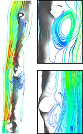
Global CSF Fluid Flow Simulation Results and Discussion: CSF flow is driven by a pulsatile waveform and Womersley (Wo) number is calculated to characterize the flow profile as in phase (<1) or lagging behind the pressure wave (>1). The Womersley number at the C4 region of the spinal SAS is Wo=4.63, T6 region Wo=4.38, and L4 region Wo=5.38. Flow at all spinal regions of interest has a phase lag behind the pressure wave. Velocity streamlines, shown in Figure 2, are
generated which visualizes recirculating flow patterns in the spinal SAS at multiple regions adjacent to nerve roots. Reynolds number is calculated at multiple regions of the spinal canal to determine if fluid is turbulent or laminar. Simulation results predict all regions of the spine to have laminar flow; C4 Re<81, T6 had Re<37, and L4 has Re<17. The presence of CSF recirculation patterns indicates micro-anatomy influence the fluid flow profiles. The pressure drop increased as the density of manually added trabeculae increased. Micro-anatomy has a significant role in the dispersion of fluid flow which will impact the distribution of drug nanoparticle complexes. Magnetic two-phasic flow predicts nanoparticle localization at regions of interest under increasing external magnetic field strength and gradient.It is plausible that the CSF pulsations and micro-anatomy are both important for preventing the formation of dead zones in the spinal SAS in which cell debris or metabolites might otherwise stagnate and even accumulate.
|
|
Figure 2: Fluid flow patterns in the spinal SAS at nerve roots. Micro-anatomy induced mixing profiles are visualized at the C4 and T7 nerves with eddies. | Figure 3: Magnetic field produced by Ne-Fe-B magnets at various locations along the CSF filled spaces in the bench top model. Magnetic field ranges from 0.051‑0.098 T (left magnet), and 0.092‑0.167 T (right magnet). Color scale visualizes the intensity of the magnetic field; white is highest, red and black lowest. |
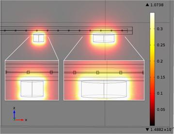
In vitro Model Results and Discussion: Multiple magentic field strengths were tested using COMSOL Multi-physics as shown in Figure 3. In the bench top model, collection efficiency at regions of interest, using a magnetic field of 0.528 T, increased with time and reached a steady collection level of 71% after 15 minutes (p-value = 0.341). No change in the magnetic retention value was observed beyond 60 minutes. The nanoparticle recovery at the region of interest increased with the rising field strength of the magnet. Percentage retentions of 48%, 61%, and 71% were observed for the surface field strengths of 0.396 T, 0.507 T, and 0.528 T respectively. Experiments utilizing the same 0.528 T magnet placed at one of the three regions of interest (C4, T6, or L4) for 15 minutes showed that location of the applied magnetic field always corresponded to the site of maximum particle retention around 76‑81%.
Therapeutic interventions for diseases of the CNS can benefit from a patient specific understanding of the effect that micro-anatomy has on the transport phenomena in the CNS leading to improved applications of novel drug delivery techniques such as usingmagnetically targeted nanocarriers.
See more of this Group/Topical: Engineering Sciences and Fundamentals