
 Google
 Yelp
 Facebook
 Twitter
 ChEnected
344104 A Platform for Retaining Native Morphology At Sub-Second Time Scales in Cryo-TEM
Introduction
Cryogenic transmission electron microscopy (cryo-TEM) is a widely used tool for morphological analysis in biology, chemistry, and polymer science. Using this technique, samples can be imaged in their native state without fixing or staining, which often cause fundamental conformational changes. Sample preparation for cryo-TEM involves removing the excess liquid deposited on a cryo-TEM grid by blotting with filter paper, which can be seen in Figure 1c-d, and plunging the grid into a cryogen (usually liquid ethane), resulting in vitrification. The blotting step is required to thin the sample, allowing the electron beam to pass through the sample, and to ensure complete sample vitrification. During this process, however, native sample morphology is compromised due to high shear rates inflicted on the sample. Additionally, species may non-specifically adsorb to the filter paper, resulting in a non-native sample concentration when imaged.
Platform Overview
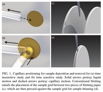 We report the development of a new sample
preparation device based on capillary action that overcomes the above
limitations. The use of a glass removal capillary aligned parallel to the grid,
as shown in Figure 1a-b, results in reduced shear and lower absorption of
particulate material from the sample. Furthermore, a deposition capillary
aligned perpendicular to the grid coupled with LabView-controlled hardware
allows for precise and sub-second resolution for time resolved studies. We
demonstrate each of the features of this platform using model samples, and
where appropriate, compare our results to those prepared using current
vitrification platforms.
Results
The efficacy of our new sample preparation technique is shown through the imaging of CTAB in phenol, Ultra Downy fabric softener, calcium carbonate intermediates, and a carbon black nanoparticle suspension. A mixture of 50 mM CTAB and 50 mM phenol, which forms wormlike micelles in aqueous solution, was utilized in order to compare the morphological differences resulting from choice of sample preparation method. The natural morphology of the wormlike micelles exhibits an entangled structure, which was captured after preparing a sample with our method. A sample prepared using a Vitrobot system with blotting and without relaxation results in wormlike micelles that are oriented parallel to each other, aligned in the direction of flow. Numerical studies were used to quantify the shear causing this alignment; it was found that shear during blotting is one to three orders of magnitude greater than for our method of parallel capillary action. To illustrate the time resolution of the system, the evolution of a mixture of aqueous calcium chloride and sodium carbonate from amorphous calcium carbonate at 700 milliseconds to calcium calcite at 5 was depicted. Another advantage of our system is the capability of imaging higher viscosity fluids. Cryo-TEM images were successfully taken of Downy fabric softener with a viscosity ~100x higher than that of water and a surface tension lower than that of water. Lastly, images of carbon black particle suspension prepared using our method showed species adsorption was reduced by ~20x compared to blotting.
Conclusion
We have developed a novel cryo-TEM sample preparation technique that allows for the vitrification of samples without the shear induced orientation of artifacts that are typical of the conventional cryo-TEM blotting sample preparation. Through the use of a glass removal capillary, we have also effectively eradicated the issue of the altering of sample nature through the adsorption of sample species typical of current preparation methods. Our technique also allows for sub-second time resolution, which is beneficial for the imaging of the intermediates of chemical or biological processes present at sub-second intervals. Our results confirm that this new sample vitrification device opens up previously unattainable regimes for sample preparation and imaging and is a powerful new tool for cryogenic transmission electron microscopy.
See more of this Group/Topical: Engineering Sciences and Fundamentals