
 Google
 Yelp
 Facebook
 Twitter
 ChEnected
344161 Droplet Microfluidic Systems For Single Cell Whole Genome Amplificiation
DROPLET Microfluidic systems FOR SINGLE CELL WHOLE GENOME AMPLIFICIATION
M. Rhee, R.J. Meagher, P. Liu, Y.K. Light, S. Yilmaz, J. Sustarich and A.K. Singh
Sandia National Laboratories, USA
The diversity of microbes in environments ranging from soil and water to the human body has been long recognized while there are big challenges for the study of microbial diversity and their roles, including extremely low cell density and uncultivable microbes. The majority of such microbial organisms are uncultivable, making it challenging to perform genome sequencing and functional characterization. Uncultured microbes in the human body may play important roles in processes related to pathologic development, and uncultured environmental microbes may also hold key metabolic pathways enabling more efficient biofuel processing and bioremediation. Even a slight change of this microbial composition may be a critical indicator of a variety of the progression of diseases; however, its complex pathway to the change is still uncertain yet. Although Metagenomics has contributed to the discovery of a great diversity of bacterial species, the complexity of microbial communities is highly beyond the tractable range of standard metagenomic techniques where relatively large amounts of starting genetic material is required. In addition, population-averaged statistical data from metagemonic study can be hardly used to identify the link between the genotype and phenotype of each individual cell. Thus, a targeted approach to identify, sort and isolate individual bacterial cells of interest from a pool of microbial samples is required to perform genome amplification and sequencing without culture. We present here droplet-based microfluidic tools to isolate individual microbial cells and amplify their whole genomes in a non-cultivation manner as a route to single-cell genome sequencing. The system consists of flow cytometry for identifying bacteria and on-demand droplet generation for cell encapsulation followed by injection of amplification reagents.
We have introduced a microfluidic platform for fluorescence in situ hybridization (FISH) on chips in our previous paper [1]. In order to identify or label specific bacteria with molecular specificity, we have used fluorescently labeled sequence of 16S rRNA for hybridization which has long been used for determining phylogenetic relationships between bacteria [2]. The following fluorescence-activated cell sorting (FACS) offers a convenient way to physically isolate individual cells. The individually isolated bacterial cell is then contained in a safe environment where crosstalk between cells or other contamination is strictly prevented. Aqueous emulsion droplets in oil phase serves as ideal containers for isolated single cells since they provide physical water/oil barriers to prevent contamination between cells. Microfluidic environments provide an ideal platform to generate and manipulate aqueous droplets in oil for various preparation steps prior to amplification. Evaporation of oil phase during long incubation time may become another challenge for successful amplification. Decrease in the amount of carrier oil due to evaporation can cause significant reduction of physical oil barriers between droplets or make the droplet exposed to atmosphere, resulting in non-specific droplet merging or breakage. To minimize evaporation during amplification, a fast and non-thermal amplification method is preferred. Multiple displacement amplification (MDA) using ϕ29 polymerase yields relatively uniform coverage when performed in a very small volume.
We present here a microfluidic processor with three modules constructed on a single chip, capable of performing (i) fluorescence-based flow cytometry and cell sorting, (ii) encapsulation of selected cells in sub-picoliter droplets, and (iii) injection of amplification reagents into the droplets for single-cell genome amplification. Especially, to our best knowledge, this is the first report in the field that proposes on-demand droplet generation for single cell sorting. Encapsulation of bacteria in picoliter plugs in particular allows us to scale down conventional assays into much smaller reaction volumes better suited to the size of an individual microbe. By dramatically reducing the reaction volume, the effective concentration of template is also increased, reducing amplification artifacts that often arise in single-cell reactions carried out at a conventional scale. Fluorescently labeled bacterial cells in the sample stream are hydrodynamically focused by two aqueous side sheath streams to minimize the detection area for more accurate reading. Multiple laser beams with desired wavelengths spot the detection point to determine the presence of desired cells using computerized PMT based on the fluorescence and scattered light. Upon the successful detection of bacterial cells, the control software triggers the microdispensing pump connected to a dispensing fluid. The dispensing fluid is injected, gets mixed with the sample stream and forcedly generates a droplet through a small orifice into the adjacent oil stream. The dispensing is programmed based on calculated delay time between detection and dispensing and the size of the droplets can be fine-tuned by dispensing pressure and pumping time. These parameters all together not only determine whether the cell or particles of interest can be encapsulated in the generated droplet or escape to the waste collection but also affect the recovery time for the next droplet generation. Typically, as the size of droplets decreases, the pump requires less recovery time. For droplets of ~50 µm in diameter, the full recovery time is approximately 200 ms, which enables droplet generation at the rate of 5 Hz. This generation rate is not comparable to that of high-throughput droplet generation systems that typically produce hundreds to thousands of droplets per second, but the on-demand nature of our system inevitably has a limitation of speed in compensation of versatile controls over droplet generation. For our microbial studies, our candidate samples of interest typically are rare and have a very low cell density, which makes this platform most suitable for the purpose. In case of high concentration samples, prior dilution will facilitate single cell encapsulation.
An encapsulated cell in the aqueous droplets is physically protected by water/oil interface. However, this physical barrier imposes another challenge of adding lysis and amplification reagents into the flowing droplets. Having a specially designed injection module down the stream, we successfully demonstrated that various reagents and buffer solutions could be merged into the cell containing droplets without losing or contaminating encapsulated cells. When the flowing droplets encounter concurrent reagent stream, by applying voltage of 100 V at the merging site, the concurrent flow injects an adjustable amount of reagents into the droplets. Because of internal circulation inside the injected droplets, almost instantaneous mixing occurs and the merged droplets become ready for the following amplification. After careful collection of droplets, the isolated single-cell genomes were amplified by multiple displacement amplification (MDA). using high purity ϕ29 DNA polymerase and their 16s rRNA genes were amplified and sequenced for identification.
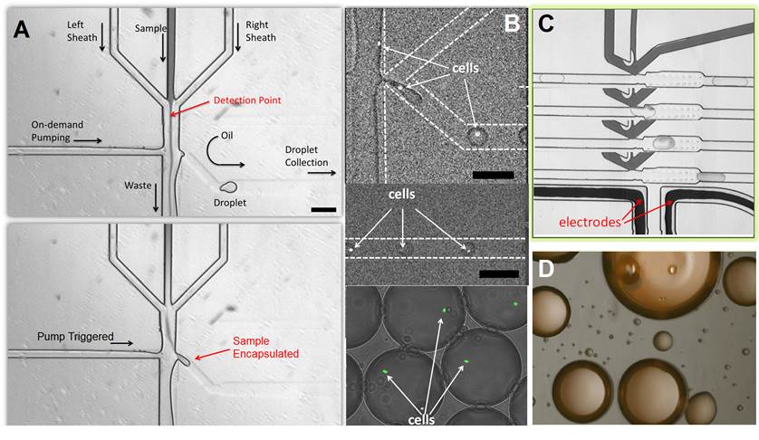
Figure Captions
A. (top) Upon the detection of bacterial cells, the control software actuates the micro-dispensing pump connected to a dispensing fluid. (bottom) The dispensing fluid is injected into the channel and forcibly reroutes the sample stream into adjacent oil stream through a small orifice, forming an emulsion droplet.
B. (top) GFP expressing E. coli cells are detected and encapsulated in aqueous droplets in oil one by one. (middle) Single E. coli cells contained in emulsion droplets flow down the channel for reagent injection. (bottom) Microdroplets serve as discrete, non-communicating containers or reaction vessels.
C. Injection of Fixed amount of reagents enabled by con-current pico-injector
D. A microscope photo of droplets containing amplified genomic DNA spread inside the droplet.
REFERENCES
[1] P. Liu et al., "Microfluidic fluorescence in situ hybridization and flow cytometry (mFlowFISH)", Lab on a Chip, vol. 11, pp. 2673-2679, 2011.
[2] R.I. Amann et al., "Combination of 16S rRNA-targeted oligonucleotide probes with flow cytometry for analyzing mixed microbial populations", Applied and Environmental Microbiology, vol. 56, pp. 1919-1925, 1990.
[3] T.D. Perroud et al., "Microfluidic-Based Cell Sorting of Francisella tularensis Infected Macrophages Using Optical Forces", Analytical Chemistry, vol. 80, pp. 6365-6372, 2008.
See more of this Group/Topical: Food, Pharmaceutical & Bioengineering Division