
 Google
 Yelp
 Facebook
 Twitter
 ChEnected
347071 A Vector-Free Antigen Delivery Platform for Cancer Vaccines
A vector-free antigen delivery platform for cancer vaccines
Authors: Armon Shareia, Shirley Maoa, Pamela A. Bastob, Gregory Szetob, George C. Hartoularosa, Siddharth Jhunjhunwalab, Darrell J. Irvineb, Robert Langera,b, Klavs F. Jensena
Affiliations: aChemical Engineering Department, bKoch Institute for Integrative Cancer Research, Massachusetts Institute of Technology. 77 Massachusetts Ave, Cambridge, MA 02139.
Introduction
Cell-based vaccines against cancer, composed of tumor antigen-loaded antigen presenting cells (APCs), hold great therapeutic potential due to their ability to activate the patient's immune system and drive a long-lasting, tumor-specific, CD8 T cell response. These therapies, such as the recently approved Provenge® for prostate cancer, have minimal side-effects relative to chemotherapies and radiation treatment. However, one of the main barriers to developing such vaccines has been achieving efficient intracellular loading of APCs with protein antigens for presentation to CD8 T cells. Since the mechanism of cross-presentation (a process wherein endocytosed proteins can induce a CD8 response) within antigen presenting cells remains elusive, one must develop a reliable method of delivering the desired antigen directly to the cell cytoplasm to advance therapies utilizing the potent effector CD8 response. In this work, our team has adapted our recently developed microfluidic delivery system (Sharei, A. et al. PNAS, 2013. 110, 1082-2087) to induce selective MHC class I antigen presentation by direct delivery of the target protein to the cell cytosol. Compared to endocytotic methods, this system has demonstrated a 10-100 fold improvement in antigen-specific CD8 T cell activation and can potentially be translated to more challenging systems, such as B cells.
Materials and Methods
This work was performed using the ovalbumin (ova) antigen presentation model. In our experiments, dendritic cells were derived from the bone marrow of healthy B6 mice and treated by the aforementioned microfluidic delivery system to facilitate intracellular delivery of the ovalbumin protein. Antibody staining, ELISA, flow cytometry, and proliferation assays using OT-I and OT-II transgenic mice were used to assess antigen presentation efficiency and T cell cytokine secretion.
Results and Discussion
In our studies, we have demonstrated enhanced MHC-I presentation of the SIINFEKL epitope by antibody staining and verified that the delivery process does not interfere with DC maturation as measured by up-regulation of CD80 and CD86 in response to stimulation by lipopolysaccharide. As illustrated in Fig. 1, this method has also successfully induced the activation/proliferation of antigen-specific CD8 T cells (derived from OT-I mice) as measured by T cell proliferation and cytokine secretion. Moreover, preliminary results indicate that this method can induce functional antigen presentation in primary B cells – a significant challenge for existing technologies.
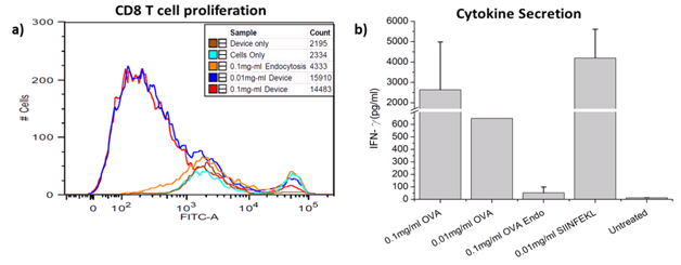
Figure 1. a) Bone marrow derived DCs were treated by the device in the presence of varying OVA concentrations and T cell proliferation was measured by CFSE staining and flow cytometry. b) Secretion of IFN-γ from CD8 T cells stimulated by treated DCs. The ‘OVA Endo' condition corresponds to an endocytosis only case.
Conclusion
The described intracellular delivery technique has demonstrated enabling potential as a platform for high efficiency antigen presentation in cancer vaccine applications.
See more of this Group/Topical: Education Division