
 Google
 Yelp
 Facebook
 Twitter
 ChEnected
463233 Dexamethasone-Releasing Polyurethane Microfiber Meshes to Facilitate Human Cardiac Stem Cell Proliferation and Differentiation
 c-kit positive human cardiac stem
cells (hCSCs) have demonstrated great
promise for treating ischemic heart failure. However, therapeutic efficiencies have
been low, likely due to limited cell survival, retention, engraftment, and
uncontrolled differentiation following transplantation. Cardiac tissue engineering has
offered new strategies to overcome these problems. Cell or biomaterial cardiac
patches, 3D constructs, and bioreactor-conditioned scaffolds have been
previously utilized to repair cardiac damage. Factors necessary to enhance cell
retention, survival rate, and ultimate cardiac differentiation include
mimicking the properties of the cardiac extracellular matrix (ECM), providing a
high surface area for display of adhesive ligands, and presenting a nano-scale
architecture with topographical features to guide cell morphology and
orientation. We
propose that elastomeric microfiber meshes that release anti-inflammatory
corticosteroids could facilitate cardiomyocyte differentiation of hCSCs, and
enhance cell retention and engraftment in vivo.
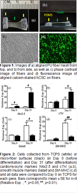
As a first step, in this study we examined aligned electrospun polyurethane (PU) microfiber meshes for engineering of cardiac patches using human cardiac stem cells (hCSCs). To this end, thin (~5 μm) elastomeric meshes consisting of aligned PU (1.7 μm diameter) electrospun micro-fibers were electrospun and suspended above glass slides using PDMS strips for supports (Figure 1). hCSCs were then seeded on these meshes and cultured for up to 31 days within differentiation media and compared to hCSC seeded in tissue culture polystyrene plates (TCPS). The viability, morphology, proliferation rate, gene, and protein expression of the hCSCs were then analyzed. Briefly, we found that cells on aligned microfiber meshes displayed an elongated morphology aligned in the direction of fiber orientation, lower proliferation rates, but increased expressions of genes and proteins majorly associated with cardiomyocyte phenotype. The early (NK2 homeobox 5, Nkx2.5) and late (cardiac troponin I, cTnI) cardiomyocyte genes were significantly increased on mesh (Nkx2.5 = 56.2 ± 13.0, cTnl = 2.9 ± 0.56,) over TCPS (Nkx2.5 = 4.2 ± 0.9, cTnI = 1.6 ± 0.5, n = 9, p<0.05 for both groups) after 31 days of differentiation (Figure 2). In contrast, expressions of smooth muscle markers Gata6 and smooth muscle myosin heavy chain (SM-MHC) were significantly decreased on microfibers. Concurrently, immunocytochemical analysis with cardiac specific antibody exhibited a similar pattern of cardiac lineage differentiation.
As the next step, we are currently incorporating the corticoid dexamethasone within the core phase of core/sheath coaxially electrospun fiber meshes and characterizing its release as a function of the thicknesses of the core and sheath phases. Our hypothesis is that the sustained release of this anti-inflammatory agent will increase hCSC proliferation and cardiomyocyte differentiation and mitigate hCSC apoptosis. Therefore, our plan is to show increased viability of doxorubicin-treated hCSCs, as well as to characterize mRNA markers of hCSC differentiation into cardiomyocytes (Nkx2.5, cTnI), smooth muscle cells (Gata6, SM-MHC), and endothelial cells (CD-31).
See more of this Group/Topical: Food, Pharmaceutical & Bioengineering Division