
 Google
 Yelp
 Facebook
 Twitter
 ChEnected
468364 Morphogen Presentation within Micro-Fiber/Collagen Composites for Ligament Tissue Engineering
 Tissue engineering holds promise in
overcoming the limitations of existing autologous and allogeneic options. However,
central to the realization of this goal is the achievement of instructive
biomaterial scaffold materials that can guide the recruitment, proliferation, and
differentiation of stem cells into organized functional tissues. In particular,
we hypothesize that scaffolds that present anisotropic topographical and mechanical
properties, as well as morphogenic signals are essential for guiding stem cell
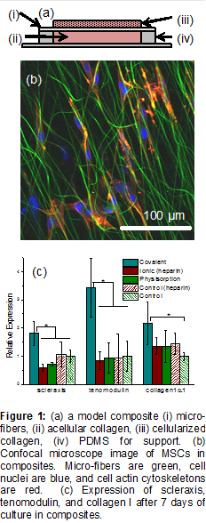
fate. To this end, we have developed model electrospun micro-fiber/collagen hydrogel
composites – consisting of thin oriented micro-fiber meshes between collagen slabs
(Figure 1a) – that allow us to selectively tune the mechanical, biochemical
properties of both the fiber and hydrogel phases and assess their effects on
stem cell proliferation and differentiation. For the goal of ligament tissue
engineering, we have previously shown that mesenchymal stem cells (MSCs) attach
to and align with the oriented micro-fibers (Figure 1b). Further, we
found that systematic variation of the micro-fiber modulus affects mRNA
expression of the tendon/ligament transcription factor scleraxis and the
contractile protein α-smooth muscle actin (not shown). Currently, we are extending
this work to determine the effect of tethering the morphogens (i.e., fibroblast
growth factor (FGF)-2 and growth and differentiation factor (GDF)-5) to the
micro-fibers.
To accomplish this, we are electrospinning thin (5-10 μm) aligned meshes of coaxial micro-fibers (~1 μm in diameter) consisting of a chitosan sheath phase and a polyurethane/polycaprolactone blend core phase on a rotating mandrel. The meshes are transferred to PDMS rings and functionalized with a mixture of FGF-2 and GDF-5 through covalent bioconjugation, ionic adsorption (to heparin-coated micro-fibers), or simple physisorption (to untreated micro-fibers). Resultant bioactive fiber meshes (1.2 cm diameter) are embedded within 0.5 wt% collagen gels containing 5×10⁴ MSCs and cultured for up to 7 days and compared to samples without morphogens. Preliminary data shows that covalent bioconjugation results in enhanced expression of ligament markers scleraxis and tenomodulin (Figure 1c). In contrast, the incorporation of morphogens onto heparin-coated and uncoated micro-fibers does not enhance expression, perhaps due to denaturation of the proteins during adsorption or subsequent desorption and loss of the proteins from the micro-fiber surfaces. Concurrently, we are examining adsorption and desorption of morphogens from planar chitosan films. Initial data indicate covalent bioconjugation of FGF-2 from a 250 ng/mL solution results in a surface concentration of 58 ng/cm2 with negligible protein loss over 7 days. In contrast adsorption of FGF-2 to chitosan and heparin-coated chitosan surfaces results in surface concentrations of 182 and 137 ng/cm2, respectively.
Our results suggest that the covalent bioconjugation of morphogens onto the micro-fiber surfaces can have a strong positive effect on stem cell fate. We postulate that the enhanced expression of ligament markers (Figure 1c) is due the localization of morphogen/receptor complexes within or near focal adhesion complexes. This co-localization may mimic the native ECM, by supporting cell adhesion, migration, and guide behavior due to integrin association with both growth factors and adhesive proteins. Ultimately, our goal is to develop thick multilayered composites suitable for implantation, where the bioactive micro-fibers present a multitude of stimuli to recruit host cells and guide tissue formation in vivo.
See more of this Group/Topical: Materials Engineering and Sciences Division