
 Google
 Yelp
 Facebook
 Twitter
 ChEnected
469419 Combined Omics Approach Reveals the Effects of a Phthalate Pollutant on Adipocyte Metabolism and Inflammation
 Introduction: Contamination
of the environment with organic pollutants has emerged as a significant public
health concern due to the pervasive nature of these contaminants.
Epidemiological studies have linked chronic endocrine disrupting chemical (EDC)
exposure to adverse effects on reproduction, development, and more recently,
metabolic diseases [1]. A growing number of studies have reported that
perinatal exposure to certain EDCs, termed obesogens,
could contribute to weight gain through an adipogenic
effect that leads to increased fat mass. To date, studies have mainly focused
on the impact of suspected obesogens on stem cell
fate and tissue development, sometimes yielding conflicting results. Less
attention has been paid to clarifying whether these chemicals can directly
disrupt metabolic regulation in differentiated cells of adult tissue. For
example, EDCs could interfere with endogenous regulatory pathways to impair the
ability of adipocytes to sequester fatty acids or activate pro-inflammatory
pathways, both hallmarks of obesity-related metabolic diseases [2]. Studies
have shown that EDCs can bind to nuclear receptors (NRs) in a variety of
tissues to disrupt the normal function of the endocrine system [3]. One such
NR, peroxisome proliferator activated receptor gamma (PPARγ),
is a particularly promising link between EDCs and their metabolic effects. This
NR recognizes structurally diverse ligands, and regulates the transcription of
genes involved in lipid storage homeostasis. Importantly, this NR interacts
with other nutrient sensors through feedback mechanisms [2], thus coupling
control over metabolism and signaling in adipocytes.
Objective: Our aim in the current work was to study the effects of low, physiologically relevant doses of EDCs on differentiated murine adipocytes, working under the hypothesis that EDCs act via both metabolic and inflammatory signaling pathways to elicit their effects.
Methods: Due to their exogenous origin, EDCs cannot be readily placed into the context of a canonical biochemical or signaling pathway. In this light, a data-driven (e.g., multi-omic) approach could provide valuable clues in determining the pathways impacted by the chemical, which in turn could lead to mechanistic insights. In this study, we combined mass spectrometry metabolomic and proteomic methods with gene expression analysis to study the biochemical changes elicited by a pervasive EDC, monoethylhexyl phthalate (MEHP), in differentiated 3T3-L1 adipocytes. To broadly assess the metabolic effects, we profiled the levels of ~50 intracellular metabolites, and performed a partial least squares discriminant analysis (PLS-DA) on the metabolite data. We also used untargeted proteomics methods to profile the levels of ~1000 cellular proteins and performed PLS-DA, pathway/network analysis, and targeted quantification on the data.
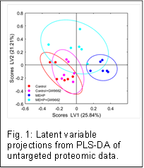
Results: To establish that EDCs elicit an inflammatory response in mature adipocytes, we screened for the effects of 3 representative EDCs - tributyltin (TBT), bisphenol A (BPA), and MEHP - by first inducing adipogenic differentiation of 3T3-L1 cells, and then treating the cells with the chemicals at doses reported in human exposure studies. At nanomolar doses, the chemicals did not affect cell morphology or fat accumulation, but significantly increased expression of inflammatory gene markers such as monocyte chemo-attractant protein 1 (MCP-1). Latent variable score projections from PLS-DA of the metabolite data showed clear differences in the metabolite profiles across the treatment groups. The differences relative to control cells treated with only the vehicle (DMSO) were most pronounced for MEHP treated cells. The corresponding loadings showed that multiple metabolites contributed to the differences, with the majority comprising free fatty acid (FFA) species. Focusing on MEHP as a potent EDC, we performed a dose response experiment to obtain a more detailed understanding of the metabolic and inflammatory responses. MEHP treatment increased the expression of an array of chemokines and cytokines, similar to the effects of the prototypical pro-inflammatory cytokine TNFα. Exposure to MEHP also altered the intracellular FFA profile, raising the levels of both saturated and unsaturated FFAs. We next performed untargeted proteomics experiments with targeted quantification to profile the impact of MEHP exposure on the levels of cellular proteins. The proteomic data indicated that the chemical exposure broadly and significantly altered the levels of enzymes and other proteins involved in regulating the lipid balance and inflammatory pathways in adipocytes. These data were consistent with our hypothesis that MEHP impacts transcriptional/translational regulation of metabolic pathways. Several of the up-regulated proteins were downstream targets of PPARγ (e.g. GPDH, FABP4), which supports the involvement of this key transcriptional regulator. Pathway enrichment analysis using the String database and Ingenuity Pathway Analysis found that lipid metabolism and PPAR signaling, respectively, were the most significantly overrepresented metabolic and signaling pathways in the discriminatory protein dataset. To further investigate the involvement of PPARγ in the observed metabolic and inflammatory effects, we tested whether inhibiting PPARγ activity would abrogate the effects of MEHP exposure. A comparison of protein expression profiles showed that co-treating the cells with the selective synthetic antagonist GW9662 during MEHP exposure attenuated MEHP-induced differences, taking the adipocytes to a phenotypic state more similar to vehicle control (Fig 1). Additionally, co-treatment with MEHP and GW9662 prevented the significant increase in chemokine and cytokine expression observed when the cells were treated with MEHP alone.
Conclusions and ongoing work: Taken together, our results support the hypothesis that the metabolic and inflammatory responses are coupled, and that PPARγ acts, at least in part, as a key mediator. Our results also support a mechanistic link between EDC exposure and adipocyte inflammation, which underpins many of the metabolic dysfunctions of obesity. The next step of this research is to apply another dimension of omics analysis, fluxomics, to better understand the metabolic alterations that lead to increased levels of FFAs, the hypothesized link between metabolic and inflammatory signaling. To this end we are repeating MEHP exposure experiments in adipocytes fed an isotopically labeled substrate, and using isotopic labeling based metabolic flux analysis to gain insight into which central carbon metabolism and lipid synthesis/breakdown pathways are perturbed. We anticipate that the results of this analysis will help guide focused loss-/gain-of-function experiments to further clarify the role PPARγ and other regulator molecules play in the observed effects.
References:
[1] Heindel (2003) Toxicol Sci 76: 247-9.
[2] Manteiga et al. (2013) Wiley Interdiscip Rev Syst Biol Med 5: 425-47.
[3] Grun et al. (2007) Rev Endocr Metab Disord 8: 161-71.
See more of this Group/Topical: Food, Pharmaceutical & Bioengineering Division