
Imaging techniques constitute a novel paradigm in characterization methods of porous media; X-ray computed tomography is of particular interest because it allows for the non-destructive determination of spatially resolved properties such as macropore space[1], and distribution of binder content[2] and minerals[3]. While the resolution is being continuously increased moving from medical scanners towards monochromatic synchrotron micro-CT and nano-CT setups, bench scale laboratory systems are still limited to voxel sizes above 1µm3, and therefore do not allow visualising micro- and meso-porosity (less than 50 nm) that is ubiquitous in both natural (e.g., clay, coal, shale) and most porous materials used in the chemical industry (e.g. adsorbent materials, catalysts, membranes). The most widespread technique for the characterization of nanoporous solids indeed is gas adsorption. While the interpretation of sub-critical adsorption isotherms in terms of pore size, pore structure and connectivity is being continuously refined[4], gas adsorption remains a technique that only provides bulk (macroscopic) information and can therefore not provide any information regarding the distribution of these properties.
The images obtained from X-ray CT scans contain spatially resolved quantitative information of the linear attenuation coefficient: the gray scale value of each voxel is the volume weighted average of the attenuation of each component and/or phase present in that specific voxel. Based on this observation, Pini[5] showed in a recent communication that it is possible to extract the excess adsorption from the subtraction of a scan of a porous solid taken in the presence of an adsorbate, and a scan of that same solid taken under vacuum or in the presence of an inert (e.g. Helium). This work has been extended here by developing the relevant experimental and analysis protocols to measure full excess adsorption isotherms by X-ray CT imaging for medical and micro-CT setups. By applying the devised protocols to fixed beds of commercial zeolite 13X pellets (ZeoChem, Uetikon Switzerland) and activated carbon rods (Norit, USA), we measured spatially distributed adsorption isotherms of CO2 -- one isotherm for each cubic millimeter -- by means of X-ray CT imaging with a medical CT scanner (Universal Systems), as illustrated in Fig. 1. Pellets can be imaged in a similar way at a larger resolution by means of micro-CT imaging (Zeiss Versa XRM500): this enables obtaining adsorption isotherms that are spatially resolved in a sub-millimeter scale.
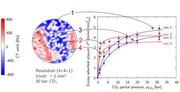
Fig. 1 X-ray CT image of a slice positioned at the interface between a Zeolite 13X and an activated carbon layer at 30 bar CO2. From difference images between CO2 scans taken at various pressures and a helium scan, excess adsorption isotherms can be reconstructed for every voxel. Two examples are shown for the rod-like particles (activated carbon, voxels 1 and 3, blue) and for the spherical particles (zeolite 13X, voxels 2 and 4, red).
Because this technique ultimately allows to quantitatively probe and visualize the spatial distribution of typical metrics, e.g. pore volume and pore size distribution, within individual pellets and across packed beds, it is anticipated that it can support the development of formulation processes of adsorbent powders into shaped particles by providing direct structural information of the properties of interest, i.e., the adsorption isotherms. Moreover, this work provides a novel way to probe nanoporosity of heterogeneous porous media in multiple dimensions and in a non-destructive way. As such, it is expected to bear significant importance in the characterization of nanoporosity within natural porous media, such as shales or reservoir rocks, where three-dimensional information on the pore size, pore volume and pore inter-connectivity are essential for the accurate description of gas transport.
References:
1. Blunt, M. J; Bijeljic, B; Dong, H; Gharbi, O; Iglauer, S; Mostaghimi, P; Paluszny, A; Pentlan, C. Pore scale imaging and modelling. Advances in Water Resources 2013, 51, 197-216.
2. Mitchell, S; Michels, N; Kunze, K; perez-Ramirez, J. Visualization of hierarchically structured zeolite bodies from macro to nano length scales. Nature Chemistry 2012, 4, 825-831.
3. Lai, P; Moulton, K; Krevor, S. Pore-scale heterogeneity in the mineral distribution and reactive surface area of porous rocks. Chemical Geology 2015, 411, 260-273.
4. Thommes, M; Kaneko, K; Neimark, A.V; Olivier, J. P; Rodriguez-Reinoso, R; Rouquerol, J; Sing, K. S. W. Physisorption of gases, with special reference to the evaluation of surface area and pore size distribution. Pure and Applied Chemistry 2015, 87, 1051-1069.
5. Pini R. Multidimensional Quantitative Imaging of Gas Adsorption in Nanoporous Solids. Langmuir 2014, 30, 10984-10989.
See more of this Group/Topical: Separations Division