
An Optically Controlled Microphysiological System for the Heart-Brain Axis
Jonathan Soucy1, Tess Torregrosa1, Abigail Koppes1, Nasim Annabi1,2, Ryan Koppes1
1Chemical Engineering, Northeastern University, Boston, MA
2Harvard-MIT Division of Health Sciences and Technology, Massachusetts Institute of Technology, Cambridge, MA Introduction:
While, there are more than 700,000 people in the United States suffer a heart attack annually, the underlying cellular mechanisms that lead to cardiac dysfunction remains poorly understood1. Previously, animal models have been used to investigate cardiovascular disease, but because of their inherent complexities, variability, and failure to translate to human physiology, in vitro alternatives must be developed2. Towards this goal, we aim to recapitulate the human cardiac sympathovagal balance in vitro to study the underlying cellular changes of cardiac tissue in response to ANS modulation.
In vivo, the heart responds to environmental changes by controlling cardiac output via the ANS. In the case of deteriorating cardiac function, the sympathetic nervous system (SNS) will innervate the cardiac tissue, while parasympathetic nervous system (PSNS) will withdraw to maintain resting heart rate3. However, as the branches of the ANS work to restore cardiac output, the overall balance of neural input leans towards the sympathetic. This understanding of the cardiac ANS pathophysiology has led to development of beta-blocker therapies to inhibit SNS activity. Conversely, the role of the PSNS in heart failure not completely understood and is an area of ongoing research. Preliminary cardiac ANS studies have shown that increased cardiac PSNS activity can reduced risk of arrhythmias, potentially leading to vagal stimulation as a new therapeutic strategy4. Yet, mimicking this imbalance in vitro for investigating the underlying cellular mechanisms has not been demonstrated.
Towards developing innervated muscle organ systems on a chip, microfluidic devices have been used for compartmentalized co-culture of skeletal muscle cells with neurons5, but only recently have applied to cardiac systems2. These neural cardiac co-culture devices can be improved upon by culturing cardiomyocytes (CM) in a physiologically relevant three-dimensional (3D) environment as well as the addition of means for real-time, bi-directional control/output of neural and cardiac function. Optogenetics allows for optical control of specific cell populations via genetic modification with light-sensitive ion channels, microbial rhodopsins6. In contrast to electrical stimulation, optogenetics enables temporally precise excitatory and inhibitory control of neural activity in distinct cell types expressing light-gated ion channels and pumps, such as channelrhodopsin 2 (ChR2) and halorhodopsin (NpHR)7. Furthermore, optogenetics is compatible with simultaneous neural recording due to the decoupling of electrical and optical signals. Independent, multiplexing modulation of multiple cell types can be accomplished with opsins possessing distinct absorption spectra such as channelrhodopsin and Chrimson8. Therefore, there is are advantages for utilizing an optogenetic toolbox to control SNS and PSNS activity independently without concern for confounding, non-specific stimulation or confounding influence on monitored electrical activity.
Here, we aim to design a custom chip to mimic an in vivo cardiac ANS and allow for optical control of neural activity. In vitro models provide numerous benefits over their in vivo counterparts by reducing variability, lowering cost, and by allowing for the inclusion of knockout transgenic cell lines used to study specific genes. Developing a functional model of the cardiac ANS will allow for an improved fundamental understanding of the relationship between the cholinergic PSNS neurons, the adrenergic SNS neurons. Additionally, by studying the cellular responses to physiological relevant changes, new therapeutic targets may be identified to treat cardiovascular disease.
Methods:
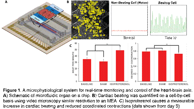
A custom microfluidic chip made of PDMS with three independent channels, separated by microposts for capillary separation of the central cardiac laden 3D hydrogel (Fig 1). This microfluidic chip will be mounted onto a 60-channel multielectrode array (MEA, Multichannel Systems) to enable both the stimulation and monitoring each cell’s electrical potential.
Pure populations of CM and cardiac fibroblast (CF) from the heart, cholinergic neurons from the intrinsic cardiac ganglia, and adrenergic neurons from the superior cervical ganglia from neonatal rat pups will be isolated for the in vitro platform. Using isolated cells, we will encapsulate varying ratios of CM and CF within a photocrosslinkable gelatin based hydrogel, gelatin methacrylate (GelMA)9 in situ using a prototype chip design and in micropatterned gel channels. We used an in-house photocrosslinking system with 405nm LEDs to polymerize GelMA in the presence of lithium phenyl-2,4,6-trimethylbenzoylphosphinate (LAP, BioBots). The use of this system resulted in CM viability significantly greater than systems using UV light; however, beating was only observed when both CM and CF were present within the gel.
Light sensitive neuron populations will be produced via transfection with adeno-associated viruses carrying either Chronos-GFP or ChrimsonR-dTomato (UNC Vector Core), to allow for independent optical control of SNS or PSNS populations. Transfection will be validated via positive expression of appropriate fluorescent reporter protein.
In addition to monitoring electrical activity, to enable quantification of cell beating activity, we developed a custom MATLAB code to calculate beating on a cell-by-cell basis using video microscopy. Furthermore, we developed a novel method to quantify the synchronicity of CM beating (Fig. 1B), based on an algorithm originally developed to investigate neuronal activity10.
Results and Discussion:
We have successfully fabricated the PDMS microfluidic platform on a 64-channel microelectrode array for real-time monitoring of both neural and muscle populations. Primary populations for cardiomyocytes, fibroblasts, as well as adrenergic, and cholinergic neurons were successfully isolated from neonatal rat pups. Independent transfection of the SNS and PSNS populations was validated with robust expression of fluorescent reporter proteins in Tyrosine Hydroxylase or Acetyl Choline positive neurons. Furthermore, seeding of our platform showed spontaneous neurite ingrowth into the cardiomyocyte laden hydrogel.
Our visual method for quantifying cardiomyocyte contraction demonstrated good correlation to contraction monitored on the MEA. Our method was able to detect changes in beating and coordinated contraction induced via the addition of Isoproterenol, a beta-adrenergic agonist (Fig. 1C). Further, using our visual quantification method of cardiomyocyte beating over long-term culture, we observed that the degree of synchrony increased to a steady value after 5 days. However, for measurements from longer culture times great than 2 weeks, there were increased arrhythmias. Nevertheless, immunocytochemistry showed that CM phenotype was maintained throughout the study.
References:
1 Loffredo, F. S., Nikolova, A. P., Pancoast, J. R. & Lee, R. T. Heart failure with preserved ejection fraction: molecular pathways of the aging myocardium. Circ. Res. 115, 97-107, (2014).
2 Oiwa, K. et al. A device for co-culturing autonomic neurons and cardiomyocytes using microfabrication techniques. Integr Biol (Camb) 8, 341-348, (2016).
3 Ripplinger, C. M., Noujaim, S. F. & Linz, D. The nervous heart. Prog. Biophys. Mol. Biol. 120, 199209, (2016).
4 Vanoli, E. et al. Vagal stimulation and prevention of sudden death in conscious dogs with a healed myocardial infarction. Circ. Res. 68, 1471-1481 (1991).
5 Uzel, S. G. et al. Microfluidic device for the formation of optically excitable, three-dimensional, compartmentalized motor units. Sci Adv 2, e1501429, (2016). 6 Deisseroth, K. Optogenetics. Nat Methods 8, 26-29, (2011).
7 Boyden, E. S., Zhang, F., Bamberg, E., Nagel, G. & Deisseroth, K. Millisecond-timescale, genetically targeted optical control of neural activity. Nat. Neurosci. 8, 1263-1268, (2005).
8 Klapoetke, N. C. et al. Independent optical excitation of distinct neural populations. Nat Methods 11, 338-346, (2014).
9 Saini, H., Navaei, A., Van Putten, A. & Nikkhah, M. 3D cardiac microtissues encapsulated with the co-culture of cardiomyocytes and cardiac fibroblasts. Adv Healthc Mater 4, 1961-1971, (2015).
10 Kreuz, T., Mulansky, M. & Bozanic, N. SPIKY: a graphical user interface for monitoring spike train synchrony. J. Neurophysiol. 113, 3432-3445, (2015).
See more of this Group/Topical: Food, Pharmaceutical & Bioengineering Division