
Gut Microbiota-derived Tryptophan Metabolites Modulate Inflammatory Response in Hepatocytes and Macrophages
Smitha Krishnan1, Yufang Ding2, Maria Choi1, Nima Saedi3, Martin L. Yarmush3, Arul Jayaraman2, and Kyongbum Lee1
1. Department of Chemical and Biological Engineering, Tufts University, Medford, MA 02155
2. Department of Biomedical Engineering, Texas A&M University, College Station, TX 77843
3. Center for Engineering in Medicine, Massachusetts General Hospital, Boston, MA 02114
The human gastrointestinal (GI) tract harbors an estimated 10 to 100 trillion microorganisms, encompassing several hundred different species. Collectively referred to as the gut microbiota, the microbes perform a number of essential physiological functions, including the metabolism of complex carbohydrates [1], development of the immune system [2], and defense against pathogens [3]. Beyond the GI tract, gut-brain, gut-lung, and gut-liver associations have also been identified, highlighting the importance of the microbiota in host physiology. Alterations in the intestinal microbiota composition (i.e. dysbiosis) have been correlated to various diseases, including obesity [4, 5], diabetes [6], and cancer [7].
Non-alcoholic
fatty liver disease (NAFLD) has emerged as the most prevalent chronic liver
disease in Western countries. It is characterized by steatosis, or fat
deposition in the liver, which can remain a benign condition. However, in
10-20% of patients, NAFLD progresses from steatosis to non-alcoholic
steatohepatitis (NASH), which can lead to cirrhosis and liver cancer. Direct,
potentially causal involvement of the microbiota in NASH was highlighted by Le
Roy et al. [11], who reported that high fat diet (HFD)-fed germ-free (GF) mice
inoculated with commensal bacteria from GI tract of HFD-fed hyperglycemic mice
developed steatohepatitis, whereas HFD-fed GF mice inoculated with bacteria
from HFD-fed, but normoglycemic mice only showed mild steatosis. This suggests
that the composition of the intestinal microbiota, and hence their metabolic
products, could determine whether NAFLD progresses to NASH. The study described
in this abstract utilizes metabolomics in conjunction with biochemical assays to
identify and characterize microbiota-dependent immunomodulatory metabolites
that could modulate liver inflammation in steatosis.
In previous
work [13], we identified a panel of aromatic amino acid (AAA)-derived
metabolites whose levels in the mouse intestine depended on the presence of the
microbiota. Furthermore, several of these metabolites were found to activate
the aryl hydrocarbon receptor (AhR), a transcriptional regulator of
host-microbiota interactions in the intestine. We compared the profiles of these
metabolites in serum, cecum, and liver samples from 14-week old male C57BL/6J
mice raised for 8 weeks either on HFD or low-fat diet (LFD). Untargeted LC-MS
experiments found that several of the previously identified gut
microbiota-dependent metabolites were significantly depleted in HFD mice
compared to LFD mice (Figure 1a-c), including Ahr activators tryptamine (TA)
and indole-3-acetate (I3A) (Figure 1d).
We next investigated the effects of TA and I3A on inflammatory responses in hepatocytes using both human (HepG2) and murine (AML12) cell lines. Cultured HepG2 and AML12 cells were treated with 10, 100, or 500 uM TA or I3A for 24 h, and then exposed to the pro-inflammatory cytokine TNF_ for 24 h in the presence of the metabolites. In both human and murine-derived hepatocytes, pretreatment with TA or I3A significantly attenuated TNF_-stimulated increases in intracellular fatty acid levels, with I3A exhibiting greater potency. Similarly, pretreatment with TA or I3A significantly attenuated TNF_Ðstimulated increases in intracellular bile acid levels. These effects were also observed when the hepatocytes were preloaded with lipid droplets prior to metabolite and cytokine treatment by growing the cells in fatty acid supplemented culture medium to mimic a steatotic state.
To confirm that the metabolites can activate the AhR pathway in hepatocytes, we applied TA and I3A to H1G1.1c3 cells expressing a stable enhanced green fluorescent protein (EGFP) reporter construct regulated by the AhR response element (AHRE) derived from the CYP1A1 promoter [14]. Consistent with our previous findings [13], 24 h exposure to TA or I3A dose-dependently increased GFP expression in the reporter cells, indicating ligand activation of the AhR in hepatocytes by these metabolites.
Finally, we investigated the effects of I3A and TA on macrophages, as polarization of resident macrophages towards a pro-inflammatory state is a key step in the progression of steatohepatitis. RAW 264.7 murine macrophages were treated with I3A and TA for 4 h, and then stimulated with palmitate for 18 h followed by LPS exposure for 6 h in the presence of the metabolites. Treatment with I3A significantly attenuated the induction of the pro-inflammatory cytokines TNFa, IL-1b and MCP-1 by palmitate and LPS in a dose dependent manner at both mRNA and protein levels. Unlike I3A, TA only decreased the gene expression of IL-1b. However, TA significantly decreased TNFa and MCP-1 expression at the protein level. Both I3A and TA significantly decreased bone marrow-derived macrophage (BMDM) migration towards MCP-1 in a dose dependent manner.
In summary, our results show that several microbiota-dependent metabolites derived from tryptophan are significantly depleted in a murine model of diet-induced liver steatosis, and that these metabolites can act directly on hepatocytes and macrophages to modulate inflammatory pathways. Our results demonstrate that these microbiota metabolites are ligands for the AhR. Taken together, our findings support the hypothesis that dysbiosis of the gut microbiota could predispose the liver to inflammation in diet-induced steatosis through an altered microbiota metabolite profile. Prospectively, insights into the mechanisms underlying the link between microbiota dysbiosis and NAFLD could provide novel strategies to treat or prevent the progression of fatty liver diseases through the use of probiotics or postbiotics.
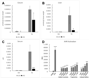
Figure 1
[1] T. Arora and R. Sharma,
"Fermentation potential of the gut microbiome: implications for energy
homeostasis and weight management," Nutrition Reviews, vol. 69, pp.
99-106, 2011.
[2] L. V. Hooper, D. R. Littman, and
A. J. Macpherson, "Interactions Between the Microbiota and the Immune
System," Science, vol. 336, pp. 1268-1273, June 8, 2012 2012.
[3] N. Kamada, G. Y. Chen, N.
Inohara, and G. Nunez, "Control of pathogens and pathobionts by the gut
microbiota," Nat Immunol, vol. 14, pp. 685-690, 2013.
[4] J. Shen, M. S. Obin, and L. Zhao,
"The gut microbiota, obesity and insulin resistance," Molecular
Aspects of Medicine, vol. 34, pp. 39-58, 2013.
[5] S. Greenblum, P. J. Turnbaugh,
and E. Borenstein, "Metagenomic systems biology of the human gut
microbiome reveals topological shifts associated with obesity and inflammatory
bowel disease," Proceedings of the National Academy of Sciences, vol. 109,
pp. 594-599, January 10, 2012 2012.
[6] J. K. Nicholson, E. Holmes, J.
Kinross, R. Burcelin, G. Gibson, W. Jia, et al., "Host-Gut Microbiota Metabolic
Interactions," Science, vol. 336, pp. 1262-1267, June 8, 2012 2012.
[7] J. C. Arthur, E. Perez-Chanona,
M. MŸhlbauer, S. Tomkovich, J. M. Uronis, T.-J. Fan, et al., "Intestinal
Inflammation Targets Cancer-Inducing Activity of the Microbiota," Science,
vol. 338, pp. 120-123, October 5, 2012 2012.
[8] R. E. Ley, F. Backhed, P.
Turnbaugh, C. A. Lozupone, R. D. Knight, and J. I. Gordon, "Obesity alters
gut microbial ecology," Proc Natl Acad Sci U S A, vol. 102, pp. 11070-5,
Aug 2 2005.
[9] P. J. Turnbaugh, R. E. Ley, M. A.
Mahowald, V. Magrini, E. R. Mardis, and J. I. Gordon, "An
obesity-associated gut microbiome with increased capacity for energy
harvest," Nature, vol. 444, pp. 1027-31, Dec 21 2006.
[10] S. G. Sheth, F. D. Gordon, and S. Chopra,
"Nonalcoholic steatohepatitis," Ann Intern Med, vol. 126, pp. 137-45,
Jan 15 1997.
[11] T. Le Roy, M. Llopis, P. Lepage, A. Bruneau,
S. Rabot, C. Bevilacqua, et al., "Intestinal microbiota determines
development of non-alcoholic fatty liver disease in mice," Gut, vol. 62,
pp. 1787-94, Dec 2013.
[12] W. R. Wikoff, A. T. Anfora, J. Liu, P. G.
Schultz, S. A. Lesley, E. C. Peters, et al., "Metabolomics analysis
reveals large effects of gut microflora on mammalian blood metabolites,"
Proceedings of the National Academy of Sciences of the United States of
America, vol. 106, pp. 3698-3703, Mar 2009.
[13] G. V. Sridharan, K. Choi, C. Klemashevich,
C. Wu, D. Prabakaran, L. B. Pan, et al., "Prediction and quantification of
bioactive microbiota metabolites in the mouse gut," Nat Commun, vol. 5, p.
5492, 2014.
[14] Nagy, Scott R., et al. "Development of a green fluorescent protein-based cell bioassay for the rapid and inexpensive detection and characterization of Ah receptor agonists." Toxicological Sciences 65.2 (2002): 200-210.
See more of this Group/Topical: Food, Pharmaceutical & Bioengineering Division