
As
articular cartilage is avascular with limited ability for
self-repair, and as a result osteoarthritis and other cartilage
injuries are biomedical burdens (Cole, Pascual-Garrido et al. 2010,
Huey, Hu et al. 2012). Current clinical and experimental treatment
methods that aim to restore articular cartilage and heal
full-thickness chondral defects are often characterized by
functionally inferior fibrocartilage, hypo-cellularity and poor
integration the repair site (Redler, Caldwell et al. 2012, Camp,
Stuart et al. 2014). Ultrasound has become an indispensable tool in
diagnostic imaging, is an FDA approved noninvasive therapy mechanism
and is used as an operative tool. While high intensity US (1-3 W/cm2)
finds utility in ablative applications, there is a paucity of
published work that investigates the ability of low intensity US
(LIUS, 1-50 mW/cm2) to improve cartilage repair outcomes and the
efficacy of LIUS to repair articular cartilage injury, in vivo,
remains unclear with conflicting outcomes (Cook, Salkeld et al. 2008,
Yang, Kuo et al. 2014, Miller 2017). Thus we investigated
low-intensity continuous ultrasound (LICUS) in cartilage restoration
(Louw, Budhiraja et al. 2013). In a departure from all previous
studies that employ low-intensity-pulsed-ultrasound (LIPUS) at 1.5MHz
and 1kHz repeat; our work has established that the cell resonant
frequency lies in the vicinity of 5 MHz for suspended and anchored
cells (Louw, Budhiraja et al. 2013, Miller 2017). Further stimulation
of chondrocytes and mesenchymal stem cells seeded in hydrogels and
macroporous scaffolds, in vitro, at the resonant frequency resulted
in enhanced chondrogenesis, improved biochemical measures,
biomechanical properties and histology scores when compared to
control, non-stimulated cultures(Guha Thakurta, Kraft et al. 2014,
Guha Thakurta, Budhiraja et al. 2015, Guha Thakurta 2016).
Successful in-vivo translation of the positive bioeffects observed in
vitro, necessitates overcoming two key challenges: (A) Identification
of an in-vivo resonant frequency and (B) Propagation of LIUS in the
joint space. Thus in order to develop a patient specific optimal
therapeutic ultrasound treatment regimen that is rigorous, the
following research objectives were undertaken: 1) the in vivo
chondrocyte resonant frequency was identified; and 2) finally the
extent of ultrasound propagation through the joint space was
theoretically modeled and verified experimentally. 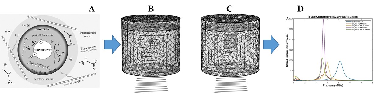

To elucidate underlying mechanisms in ultrasound induced bioeffects, we first showed that low-intensity ultrasound (LICUS) applied at resonance (~ 5MHz) induces deformation equivalent to that applied at 1MHz and significantly higher intensities (170kPa) (Miller 2017). The stored mechanical energy as a result of this deformation was maximized at resonance and the energy density in the nucleus was twice as high as in the cytoplasm. A mechanochemical model that linked the mechanical stimulation of ultrasound and the increased mechanical energy density in the nucleus to the downstream targets of the ERK pathway showed that ultrasound stimulation induces frequency dependent gene expression as a result of altered rates of transcription factors binding to chromatin. The bifurcation behavior of the cell when it is excited near resonance showed multiplicity. At positive detuning the mechanical energy coupled to the cell is small, it is higher at resonance but significantly higher at sub-resonant frequencies in the multiplicity range. Thus, there exists an optimal range of frequencies for ultrasound treatment where mechanical energy coupling is maximized (Miller 2017). As the resonant frequency is highly dependent on the mass and stiffness, we identified the resonant frequency of an in vivo chondrocyte to be in the range of 3.5−4.1 𝑀𝐻𝑧 (Fig. 1D).
Limited research has been done to determine the extent of US propagation through the joint space. Only reported study we could find is the one by White and coworkers where they experimentally investigated the propagation of a 1.5 MHz frequency pulsed US regimen through the joint space of a cadaver human knee (White, Evans et al. 2007, White, Evans et al. 2010) and did not explore the use of higher frequencies. However, their model did not include the presence of tissue, cartilage, and meniscus to allow a better match to their experimental work. Due to greater attenuation in tissues rich in protein and the use of water, their model would overestimate the field strength (Fig 2. bottom black line). Thus, there is a need to develop a clinically translatable US regimen that takes into account properties of the cell and joint tissue. We investigated the extent of ultrasound propagation through the rabbit knee joint space and the sheep elbow space and coupled these experimental results with computer simulations of the joint spaces. The joint space leads to high attenuation thus the aid of computational models can assist clinicians in determining the effectiveness of ultrasound on patient specific cartilage restoration and optimal transducer positioning.


See more of this Group/Topical: Topical Conference: Chemical Engineers in Medicine