
Zhenguo Gao 1,2 Dan Zhu 1,2 Yuanyi Wu 1 Sohrab Rohani 1* Junbo Gong 2, Jingkang Wang 2
1. The University of Western Ontario, Department of Chemical and Biochemical Engineering, London, Ontario N6A 5B9, Canada
2. Tianjin University, School of Chemical Engineering and Technology, Tianjin, 300072, P. R. China
Keywords: Ultrasonic irradiation, Nucleation, Motion-based multiple object tracking <>Introduction
Nucleation from solution has aroused great interest over the past decades. Scientists have considered atomistic levels on the order of 10-10 m and time scales in the order of 10-13 s to unravel the nucleation mechanism with the help of advanced instruments1 . However, in industrial crystallization, there is still a limitation in monitoring nucleation at the initial stage of critical nucleus formation2 . Existing measuring techniques used to monitor nucleation include the focused beam reflection measurement (FBRM), turbidity, ultrasonic velocity, electrical conductivity and light transmittance measurement of bulk solution3 . The accuracy of the monitoring devices affects the measurement of nucleation kinetics. In order to improve the accuracy, progress has been made based on high-speed imaging, multivariate statistical process monitoring charts, etc4,5 . Recently, motion-based multiple object tracking (MMOT) has been widely used in computer vision field. The MMOT method detects moving objects in a video stream, predicts their locations in the next frame, and records the object count automatically, which shows its potential application in monitoring the occurrence and disappearance of nuclei in a bulk solution.
External factors such as ultrasound, microwave, magnetic field and electric field have been studied to control crystal nucleation, which exhibit effectiveness in some cases6,7 . Ultrasonic irradiation creates sequential compression and expansion of the solution, which leads to bubble formation and growth. Finally, the bubbles collapse and release energy, promoting nucleation within a short induction time and at a lower supersaturation level. Parameters, e.g. ultrasound power, frequency, with or without pulse, have significant influence on nucleation process and crystal qualities8,9 . In this study, the MMOT was introduced in the crystallization area, for the first time, to monitor the nucleation of L-glutamic acid. Results showed that ultrasonic irradiation shortened the induction time dramatically. The MMOT-based device showed higher accuracy and precision compared to the FBRM technology as the bubbles were transparent in the MMOT technology, but considered as nuclei by the FBRM. <>Materials and Experiments
L-glutamic acid (LGA) was used as model compound. An ultrasonic processor was inserted into a double jacketed crystallizer to generate ultrasound. During nucleation process, a waterproof USB-based micro-camera (Magnification: 1-300X) was used to record a video stream to monitor the nucleation process as shown in Figure 1(a). A home-designed vial adaptor with a solution flow channel (Figure 1b) was printed by a 3D printer and connected to the end of the micro-camera to allow capture crystal images as the slurry flowed through the adaptor. Figure 2 shows the experimental setup. In the first step, clear solutions were prepared in the crystallizer at 80℃. Afterwards, nucleation temperature was reached as soon as possible by switching the 3-way valve to the cooling refrigerated circulator. The temperature was kept constant to study the nucleation kinetics until the crystals appeared in the video stream. The images recorded by micro-camera were parsed by MMOT in MATLAB.
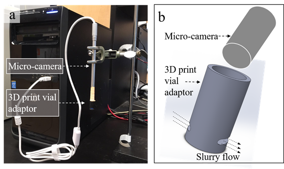
Figure 1. (a) Portable USB-based micro-camera setup; (b) 3D print vial adaptor.
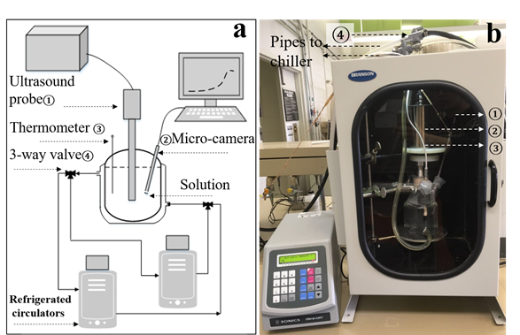
Figure 2. (a) Schematic of experimental setup; (b) Experimental setup picture. Results and Discussions
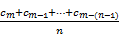
Different conditions of solution concentration (30 g/L and 40 g/L) and nucleation temperature (35℃ and 45℃) were tested. The tracking model is solely based on motion, in which the background is subtracted and a Kalman filter is used to predict the assigned track’s location in next frame. The nuclei count in MMOT was smoothed by a moving average (MA) model as shown in equations 1 and 2.
MA =
 =
 (1)
MA
current = MA prev +  (2)
Where c indicates the nuclei counts along the time series and MA prev is the previous averaged value. The moving window n was optimized to be 30 which tightly fitted the nuclei counts. The time at which a continuous increase in the micro-camera counts was sensed was taken as the onset of nucleation. The induction time was calculated as the period between the time the ultrasound processor was switched on and the onset of nuclei occurrence. Under the ultrasonic irradiation at 40 g/L and 35℃, Figure 3 and Figure 4 show the results at 38 s and 95 s of same process after the ultrasonic processor was started.
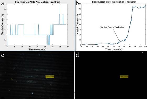
Figure 3. Detection results at 38 s with ultrasonic irradiation (14 W). (a) In-situ plot of nuclei counts versus time based on MMOT; (b) Plot of nuclei counts against time of the whole process. The blue line and black line are raw data and smoothed data. (c) Detected objects marked in an original video. (d) Detected objects marked in the video after background subtraction.
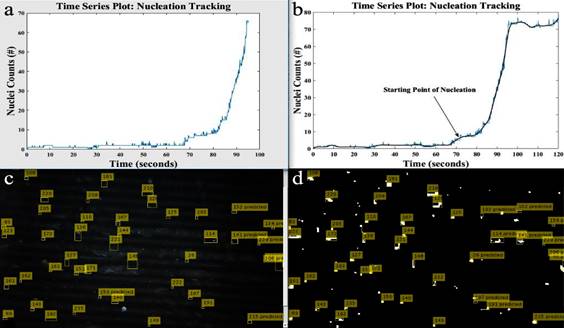
Figure 4. Detection results at 95 s with ultrasonic irradiation (14 W). (a) In-situ plot of nuclei counts versus time based on MMOT; (b) Plot of nuclei counts against time of the whole process. The blue line and black line are raw data and smoothed data. (c) Detected objects marked in an original video. (d) Detected objects marked in the video after background subtraction.
As shown in Figure 3c and 4c, only one object had been detected, which indicates no nucleation occurred before 38 s, while large quantities of objects had been detected after 95 s. A continuous increase of nuclei counts was detected after 72 s in Figure 4a, with the constant nucleation occurring in raw image (Figure 4c), which confirmed the onset of nucleation at 72 s. Comparing Figure 4a with Figure 3a, it can be concluded that the threshold of background noise is around 5, which can be ignored before the continuous increase of nuclei count. At least six experiments were repeated under the same condition and average results were reported in Table 1.
Table 1. Comparison of induction time and crystal form of LGA with/without ultrasonic irradiation.
Ultrasonic power (W) | Concentration (g/L) | Nucleation temperature (℃) | S0, α (C0/C*) | S0, β (C0/C*) | Induction time (s) | Standard deviation (s) | Crystal form |
14 | 30 | 35 | 1.91 | 2.51 | 181.3 | 8.2209 | |
14 | 40 | 35 | 2.54 | 3.35 | 77.8 | 4.0249 | |
14 | 30 | 45 | 1.31 | 1.78 | 585.6 | 22.1088 | α, β |
40 | 45 | 1.74 | 2.37 | 113.0 | 7.7136 | α, β | |
0 | 30 | 35 | 1.91 | 2.51 | 1824.3 | 100.8806 | α |
0 | 40 | 35 | 2.54 | 3.35 | 381.3 | 40.8084 | α |
0 | 30 | 45 | 1.31 | 1.78 | 3600.0 | 169.7056 | β |
0 | 40 | 45 | 1.74 | 2.37 | 737.3 | 68.3002 | β |
Note: Standard deviation was calculated by 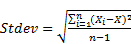 , and X
means average of all
 (i=1, 2, 3…, n).
There is a sharp decrease in induction time compared with the nucleation kinetics without ultrasonic irradiation 10. For a rising of nucleation temperature, the induction time is longer. Ultrasonic irradiation shows potential application to promote nucleation in crystallization process design. Experimental results show the measurement accuracy based on MMOT is much better than FBRM as the latter does not distinguish between the nuclei and the bubbles generated by ultrasound. However, limitation exists in MMOT method, which is similar to the PVM (Particle Vision and Measurement) technology, the detection limit of objects count is around 100 and the occurrence of multiple crystals can affect the accuracy. Conclusions
In this study, the MMOT method was, for the first time, introduced in the crystallization field to monitor nucleation process. Results show better accuracy compared with other methods such as the FBRM technology. The nuclei count measured by MMOT, which is different from the FBRM count, can be used as a parameter to control nucleation in designing crystallization processes. Based on the MMOT technology, potential applications can be developed to measure solubility, metastable zone width, particle size and even solution-mediated polymorphic transformation. <>Reference
1. J. Zhang, P. Chen, B. Yuan, W. Ji, Z. Cheng, and X. Qiu, Science, 2013, 342, 611–4.
2. A. S. Myerson, Handbook of industry crystallization, Butterworth-Heinemann, 2002.
3. C. Brandel and J. Horst, Faraday discussions, 2015, 179, 199–214.
4. S. Bauerecker, P. Ulbig, V. Buch, L. Vrbka, and P. Jungwirth, J Phys Chem C, 2008, 112, 7631–7636.
5. L. Simon, K. Oucherif, Z. Nagy, and K. Hungerbuhler, Chem Eng Sci, 2010, 65, 4983–4995.
6. S. Jiang, University of Leeds, 2012.
7. D.-C. Yin, Prog Cryst Growth Ch, 2015, 61, 1–26.
8. S. Bhangu, M. Ashokkumar, and J. Lee, Cryst Growth Des, 2016, 16, 1934–1941.
9. M. Jiang, C. Papageorgiou, J. Waetzig, A. Hardy, M. Langston, and R. Braatz, Cryst Growth Des, 2015, 15, 2486–2492.
10. Schöll, Vicum, Müller, and Mazzotti, Chem Eng Technology, 2006, 29, 257–264.
See more of this Group/Topical: Separations Division