
Here we present preliminary results towards the characterization of the dielectrophoretic spectrum of different Candida cells. In the last two decades, Candida infection has emerged as the fourth most common cause for morbidity and mortality in hospitalized patients in the United States [1]. Five Candida species have been found to be responsible for ninety percent of the all the cases of Candida infection: Candida albicans, Candida glabrata, Candida tropicalis, Candida krusei, and Candida parapsilosis [2]. The current diagnosis of Candida infection begins with the culturing of tens of milliliters of blood from the patient, which usually takes several days for detection. During the time waiting for culture results, infection may become progressively worse, leading to complications in treatment or death. These complications can be exacerbated by beginning treatment regimens that may not be effective for the particular Candida species causing the infection. Hence, there is a need for rapid, species-specific detection of infection from Candida cells. Dielectrophoresis (DEP) is a promising technique for the isolation and detection of the Candida cells within a few hours. Therefore, it is important to characterize the DEP spectrum of these cells to understand how DEP parameters (i.e. frequency, voltage, signal waveform) may contribute to the degree to which cells can be trapped and separated for detection. To date, we have characterized the frequency dependent behavior of Candida albicans, Candida tropicalis, and Candida parapsilosis.
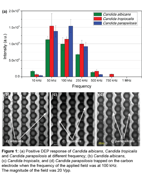
Strains of C. albicans, C. tropicalis, and C. parapsilosis were cultured in Yeast Malt Broth (YMB) supplemented with a 40% glucose solution at a 1:100 glucose to YMB ratio. Citric Acid was added to the YMB media to adjust the pH to 3.5. Cells were grown in suspension and dynamic incubation (shaking at 215 rpm) at 28°C for two days. For DEP experiments, the experimental media was prepared for cell suspension and buffer solution by dissolving 15% sucrose, 0.3% dextrose, and 0.1% bovine serum albumin (BSA) in DI water. The media conductivity was 12.62 µS/cm. We prepared the experimental cell suspension by suspending 300 µl of the cell culture in 5 ml of the experimental media followed by washing and re-suspending in experimental media. The cell concentrations of the cell suspensions were in the order of 107 cells/ml. The fabrication of the carbon-electrode DEP device has been detailed by our group in previous publications[3]. We flowed 58 µl of cell suspension through the electric field of a sinusoidal signal of constant 20 Vpp magnitude and varying frequencies ranging from 10 kHz to 1 MHz. 100 µl of clean buffer was flowed through the DEP chip at a constant flow rate of 10 µl/min to wash any trapped cells. We recorded the release of the cells at the end of the electrode arrays upon turning off the signal. Subsequent image analysis was completed with ImageJ, an image-processing software, to measure the average intensity throughout the cell elution. The results from the image analysis (Figure 1a) show the positive DEP response of cells with different frequency. Figure 1b, 1c and 1d shows the C. albicans, C. tropicalis and C. parapsilosis cells trapped on the carbon electrodes under positive DEP, respectively.
Ongoing work includes characterizing the DEP spectrum of C. glabrata and C. krusei. We intend to find their cell-wall capacitance from these DEP spectra so that they can be used in any numerical study involved the Candida cells. Future work includes selectively isolating and concentrating any Candida species from other Candida species using DEP.
References:
ADDIN Mendeley Bibliography CSL_BIBLIOGRAPHY [1] D.R. Andes, N. Safdar, J.W. Baddley, G. Playford, A.C. Reboli, J.H. Rex, J.D. Sobel, P.G. Pappas, B.J. Kullberg, Impact of treatment strategy on outcomes in patients with candidemia and other forms of invasive candidiasis: A patient-level quantitative review of randomized trials, Clin. Infect. Dis. 54 (2012) 1110–1122. doi:10.1093/cid/cis021.
[2] S. Chakravarthi, N. Haleagrahara, A comprehensive review of the occurance and management of systematic candidiasis as an opportunistic infection, Microbiol. J. 1 (2011) 1–7. doi:10.3923/mj.2011.1.7.
[3] M. Islam, R. Natu, M.F. Larraga-Martinez, R. Martinez-Duarte, Enrichment of diluted cell populations from large sample volumes using 3D carbon-electrode dielectrophoresis, Biomicrofluidics. 10 (2016) 033107. doi:10.1063/1.4954310.
See more of this Group/Topical: 2017 Annual Meeting of the AES Electrophoresis Society