
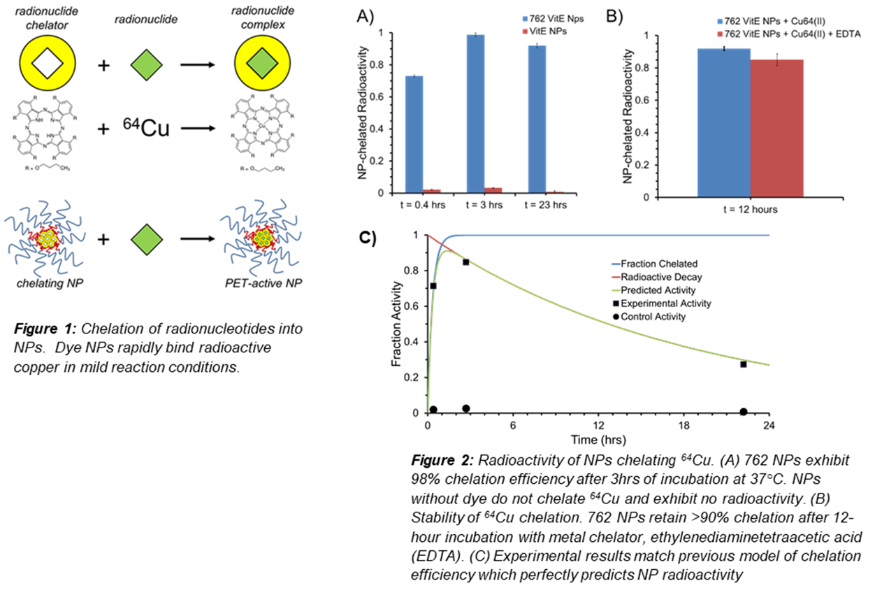
Early detection of tumors is key to the success of clinical intervention and patient outcomes. Thus, there is strong demand to develop diagnostic technologies that can detect Alzheimer’s, infections, and cancers at earlier stages when the disease is most manageable. Positron emission tomography (PET) is currently among the most sensitive imaging modalities for cancer diagnostics but require radionucleotide localization to targeted areas. Nanoparticle (NP) contrast agents can drastically increase local concentrations of radionuclides, lowering the detection limit and increasing the sensitively of PET imaging to enable detection of small and early-stage cancers that would otherwise be overlooked. However, previous methods to load NPs with PET radionuclides for in vivo imaging require harsh loading conditions or NP surface modifications that alter or diminish targeting abilities. We here present the technology for encapsulating phthalocyanine dyes inside water dispersible and polyethylene coated (PEG) NPs, using the self-assembly technique Flash Nanoprecipitation. Under the very mild conditions at pH6 and 37 C, these phthalocyanine NPs rapidly and spontaneously chelate metals, and can act as sinks for PET radionuclides, such as 64Cu, to produce PET-active NPs (Fig. 1). By measuring the absorbance shifts of the encapsulated dye over time, we developed a reaction rate model that can be used to predict chelation rates of non-radioactive ‘cold’ metals with given reaction parameters. This model was then verified with radioactive ‘hot’ metals, where chelation efficiency could be assessed directly by measuring NP radioactivity. We demonstrated that, within three hours, 762 NPs could chelate radioactive 64Cu with 98% efficiency, and retained > 90% activity even after incubation with ethylenediaminetetraacetic acid for 12 hours (Fig. 2). This result matched perfectly with the previous reaction rate model and demonstrates the use of dye absorbance shifts to accurately predict and optimize chelation reaction conditions, reducing the number of radioactive procedures required. This development, optimization, and verification of a rapid and facile radionuclide labeling method promises to increase sensitivity and expand applications of NP-based PET imaging.
See more of this Group/Topical: Particle Technology Forum