
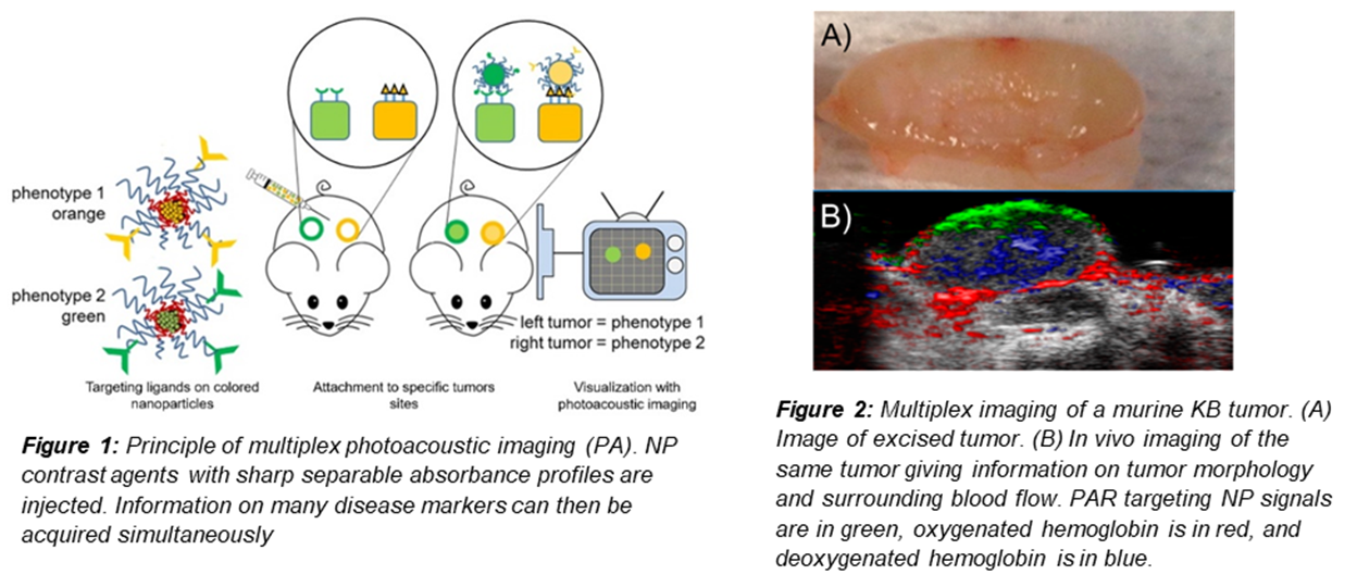
Modern cancer therapeutics have shifted focus from generic therapeutics to developing personalized treatment plans for greater safety and effectiveness. Since drugs are rapidly being developed specifically for each cancer phenotype, there is an increased demand for fast and inexpensive methods to determine cancer phenotypes and morphologies. Current diagnostic imaging modalities utilizing X-ray CT, MRI, and PET scans are limited to black-white images that cannot be used to differentiate multiple disease marker contrast agents at a time. In addition, the high costs of capital equipment and dangers of excessive imaging associated with these techniques inhibit their widespread use in diagnostics. Photoacoustic (PA) imaging, a hybrid light and sounds imaging technique, has shown to be a safe and inexpensive diagnostic technique with high spatial resolution in 3D. Traditional PA contrast agents, however, tend to have broad absorption peaks in the NIR range which renders it difficult to simultaneously image more than one signal at a time in deep tissue. Here we present the formulation of a series of PA active NPs with sharp and separable absorbance profiles in the NIR range for simultaneous multiplexed imaging (Fig. 1). PA dyes are encapsulated inside NPs using the controlled self-assembly mechanism, Flash Nanoprecipitation (FNP). Four new contrast agents, with sharp absorbance maxima between 600-900 nm, were created by encapsulating a variety of phthalocyanine derivatives. We were able to concurrently detect the concentrations of contrast agents mixed together with >95% deconvolution efficacy. Using these dye particles, we demonstrated that information on tumor morphology and surrounding blood flow can be acquired simultaneously (Fig. 2). As proof of concept, we co-injected RGD modified NPs and non-modified NPs with different labeling agents and tracked NP biodistributions for both particles simultaneously. Using this technology, we accessed the effect of NP ligand modification on both targeting efficacy onto the tumors and off-targeting accumulation in the liver using a single animal model. Over modification of the NPs resulted in rapid liver clearance and poor accumulation in the tumor; at low modifications, the tumor to liver accumulation ratio is 9.9 ± 4.2, while at high RGD modifications the tumor to liver accumulation ratio is 52 ± 22. Our particles enable new quantitative and rapid methods of assessing nanoparticle biodistributions.
See more of this Group/Topical: Topical Conference: Chemical Engineers in Medicine