
Multivalent soluble antigen arrays target antigen presenting B cells and dampen antigen-specific signaling to promote therapeutic efficacy in multiple sclerosis
Brittany L. Hartwell, Chad J. Pickens, Martin Leon, Laura Northrup, Francisco Martinez-Becerra, Cory Berkland
The University of Kansas
Many current therapies for autoimmune diseases such as multiple sclerosis (MS) act through nonspecific targeting of the immune response, rendering global immunosuppression, poor efficacy, and adverse side effects. To address a pressing need for safer and more effective therapies, we developed multivalent soluble antigen arrays (cSAgAs) to induce antigen-specific tolerance in MS. cSAgAPLP:LABL consists of a flexible hyaluronic acid (HA) linear polymer backbone click-conjugated with multiple copies of autoantigen (PLP) and adhesion inhibitor (LABL) peptides. We hypothesized that multivalent presentation of both peptides would enable antigen-specific targeting and enhanced binding with autoreactive antigen-presenting cells (APCs), thereby interrupting signaling between APCs and T cells to halt propagation of an autoimmune response. In vivo studies established that cSAgAPLP:LABL is therapeutically effective against experimental autoimmune encephalomyelitis (EAE), a murine model of relapsing-remitting MS. To elucidate the moleculeÕs therapeutic cellular mechanism, flow cytometry binding and calcium flux signaling assays were performed alongside live cell imaging to evaluate binding, specificity, and signaling modulation in different cell populations.
Studies performed in vitro in a model B cell system revealed that cSAgAPLP:LABL exhibited enhanced binding over polymer alone (cHA) and polymer conjugated with PLP only (cHAPLP) or LABL only (cHALABL). Furthermore, cSAgAPLP:LABL exhibited high avidity antigen-specific binding, targeting the B cell receptor (BCR) to dampen BCR-mediated calcium signaling. These observations suggested the moleculeÕs action at the cell surface led to B cell anergy, a state of antigen unresponsiveness induced by continuous BCR binding and marked by reduced calcium flux signaling.
cSAgAPLP:LABL binding and signaling were then evaluated ex vivo in mixed primary splenocytes isolated from EAE and healthy mice to determine which immune cell subsets were targeted and whether this resulted in an anergic phenotype. Flow cytometry binding studies were performed in splenocytes antibody-labeled for B cells (CD19+), T cells (CD3+), dendritic cells (CD11c+), and autoimmune-associated B cells (CD19+CD11c+, ABCs), a subclass of B cells that act as potent antigen presenting cells in the spleen during autoimmune disease. cSAgAPLP:LABL exhibited significantly higher binding in B cells, particularly ABCs, over other cell types (Figure 1AB). Additionally, B cell binding was significantly higher in EAE cells than healthy cells, suggesting disease-specific targeting that was not present in T cells (Figure 1CD). These results support our hypothesis that cSAgAPLP:LABL targets B cells and APCs over other cell types in a disease-specific manner.
In addition to reduced calcium flux signaling, another result of B cell anergy is down-regulation of costimulatory signals (i.e., CD80 and CD86) whose presentation is essential for autoreactive T cell activation. Thus, we investigated CD80/CD86 expression in splenocytes under two conditions: (1) harvested from EAE mice at peak of disease and treated in vitro during simultaneous PLP antigen challenge to evaluate the effect on costimulatory signaling, and (2) harvested from mice treated in vivo and re-challenged with PLP antigen ex vivo to evaluate lasting tolerance. First, in vitro treatment with cSAgAPLP:LABL caused significant down-regulation of CD86 in EAE splenocytes harvested from mice at peak of disease, causing B cells and ABCs to signal more like healthy cells rather than activated APCs (Figure 2AB). Furthermore, B cells and ABCs from mice treated with cSAgAPLP:LABL in vivo (Figure 2C) did not respond to ex vivo PLP rechallenge with a significant increase in CD80/CD86 expression, again resembling healthy cells (Figure 2DE). Splenocytes from the treated mice also exhibited elevated levels of regulatory cytokines IL-10 and IL-4. Combined, these results indicate that treatment with cSAgAPLP:LABL achieved efficacy by inducing PLP-specific anergy in B cells, resulting in reduced APC capacity and lasting tolerance to autoantigen. We conclude that cSAgAPLP:LABL offers a promising option for antigen-specific immunotherapy to repress autoimmune disease.
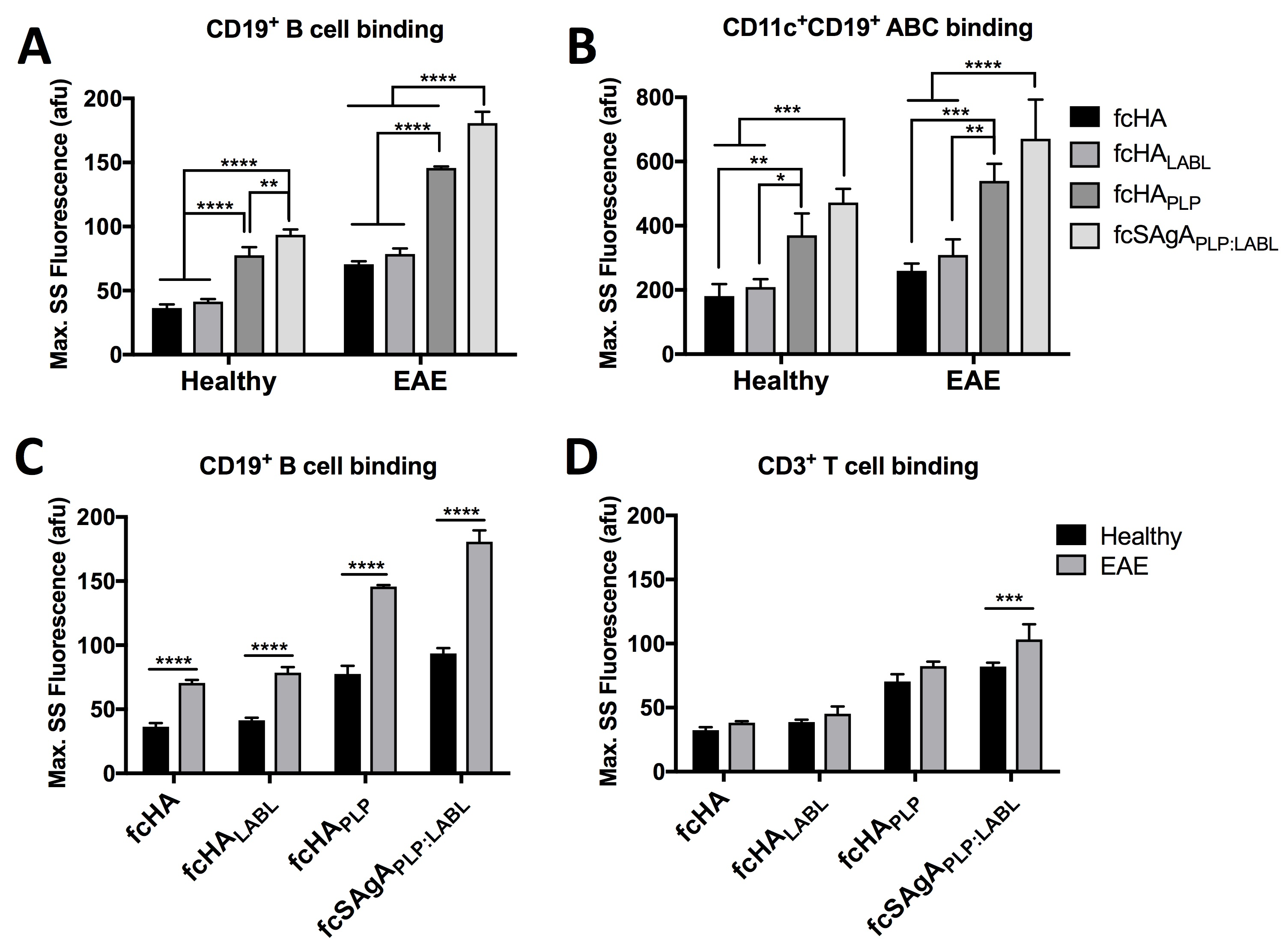
Figure 1. Maximum steady state (max. SS) binding in antibody-labeled splenocytes isolated from EAE and healthy mice determined by flow cytometry, comparing binding of fluorescently-labeled molecules in (A) CD19+ B cells, (B) CD19+CD11c+ autoimmune-associated B cells (ABCs), (C) CD19+ B cells, and (D) CD3+ T cells. Splenocytes were isolated from mice at peak of disease and cultured for 72 hr prior to the experiment. Statistical significance was determined by ANOVA followed by TukeyÕs post hoc test with p<0.05 and n=3 (*p<0.05, **p<0.01, ***p<0.001, ****p<0.0001).
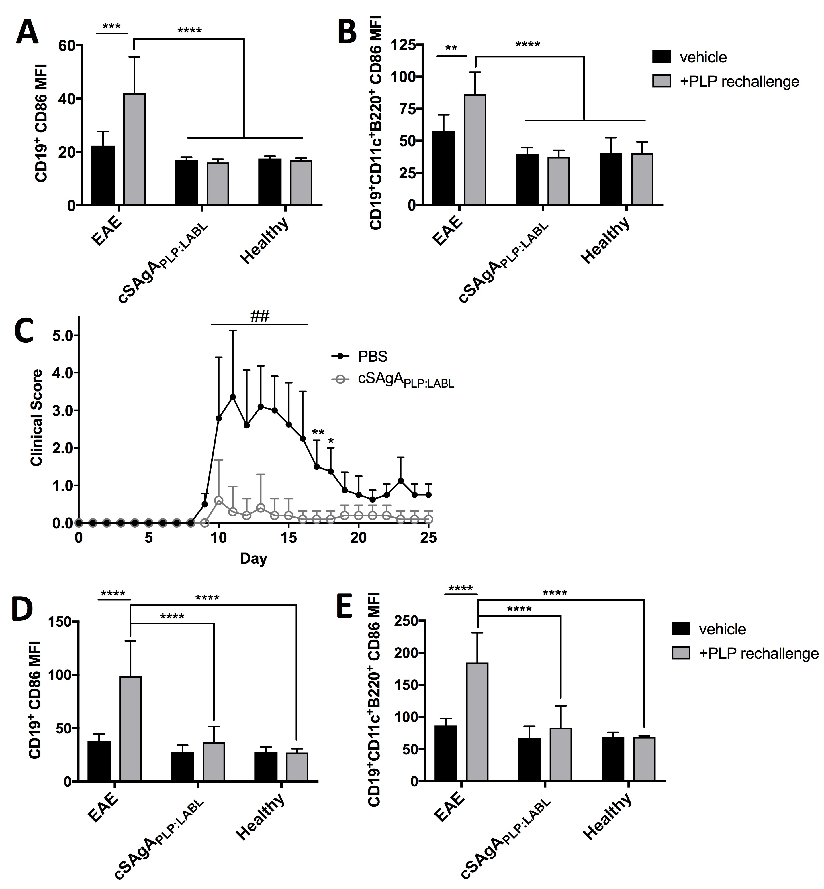
Figure 2. Costimulatory marker expression was evaluated in antibody-labeled EAE splenocytes following in vitro or in vivo treatment with cSAgAPLP:LABL, compared to EAE and healthy controls. (A/B) Splenocytes isolated from EAE mice at peak of disease were treated with cSAgAPLP:LABL for 72 hr in vitro in the presence or absence of PLP. CD86 expression (anti-CD86 mean fluorescence intensity, MFI) was evaluated by flow cytometry in (A) CD19+ B cells and (B) CD19+CD11c+B220+ autoimmune-associated B cells (ABCs) to evaluate APC costimulatory signaling. (C) EAE mice were treated in vivo with cSAgAPLP:LABL versus PBS negative control on days 4, 7, and 10, where higher clinical score indicated greater disease severity (n=5). (D/E) Splenocytes isolated from mice on day 25 following in vivo treatment in (C) were rechallenged with/without PLP for 72 hr. CD86 expression was evaluated by flow cytometry in (C) CD19+ B cells and (D) CD19+CD11c+B220+ autoimmune-associated B cells (ABCs) to evaluate lasting tolerance to autoantigen. Statistical significance (compared to the negative PBS control) was determined by ANOVA followed by TukeyÕs post hoc test with p<0.05 and n=3 (*p<0.05, **p<0.01, ***p<0.001, ##/****p<0.0001).
See more of this Group/Topical: Materials Engineering and Sciences Division