
Nanoparticles made by aerosol-based technologies find a wide range of promising applications (Pratsinis, 2010), though concerns for their potential health impact exist (Xia et al, 2009). Upon in vitro cytotoxicity evaluations of such particles, agglomeration and settling need to be considered as they influence directly the particle dose that is deposited on the employed cell cultures (Limbach et al, 2005).
Here, nanoparticles of various chemical compositions and sizes are made by flame spray pyrolysis (Madler et al, 2002) and their in vitro cytotoxicity against murine macrophages is evaluated (Sotiriou et al, 2014), accounting for their agglomeration and settling in cell culture media. The agglomerate size distributions of the particles in suspension are measured by dynamic light scattering (Wengeler et al, 2006) and their settling is evaluated by ultraviolet-visible spectroscopy (Sotiriou and Pratsinis, 2010). The cell viability after 24 h incubation with such particle suspensions is monitored by a tetrazolium salt reduction assay in both upright (Sotiriou et al, 2014) and inverted cell culture configurations (Cho et al, 2011, Lee et al, 2014) (Figure 1).
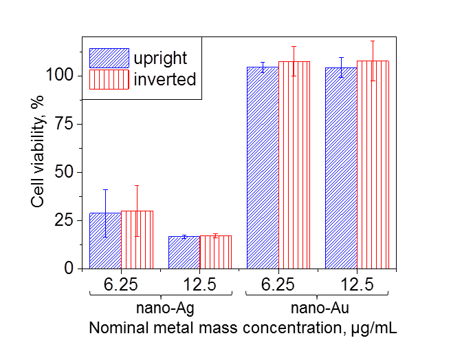
Figure 1. Relative viability of RAW 264.7 murine macrophages after 24 h incubation with nanosilver or nanogold suspensions as a function of nominal metal mass concentration in standard upright and inverted cell culture configurations.
For given nominal particle mass concentrations, three categories of nanoparticles can be distinguished, based on their cytotoxicity. Materials such as gold induce no reduction in cell viability over a wide concentration range (Sotiriou et al, 2014) and independent of the spatial configuration of the cells (Figure 1). Materials such as silver particles of relatively small crystal size (~8 nm) induce significant cytotoxicity in both configurations due to silver ion release from their surface (Sotiriou et al, 2014) (Figure 1). Finally, materials such as agglomerated silica particles induce higher cytotoxicity in upright than in inverted cell culture configuration, due to enhanced particle-cell contact in the former.
Cho, E.C., Zhang, Q. and Xia, Y.N. (2011) Nat. Nanotech. 6, 385-391.
Lee, S.H., Moroz, E., Castagner, B. and Leroux, J.C. (2014) J. Am. Chem. Soc. 136, 12868-12871.
Limbach, L.K., Li, Y.C., Grass, R.N., Brunner, T.J., Hintermann, M.A., Muller, M., Gunther, D. and Stark, W.J. (2005) Environ. Sci. Technol. 39, 9370-9376.
Madler, L., Kammler, H.K., Mueller, R. and Pratsinis, S.E. (2002) J. Aerosol Sci. 33, 369-389.
Pratsinis, S.E. (2010) AIChe Journal 56, 3028-3035.
Sotiriou, G.A., Etterlin, G.D., Spyrogianni, A., Krumeich, F., Leroux, J.-C. and Pratsinis, S.E. (2014) Chem. Commun. 50, 13559-13562.
Sotiriou, G.A. and Pratsinis, S.E. (2010) Environ. Sci. Technol. 44, 5649-5654.
Wengeler, R., Teleki, A., Vetter, M., Pratsinis, S.E. and Nirschl, H. (2006) Langmuir 22, 4928-4935.
Xia, T., Li, N. and Nel, A.E. (2009) Annu. Rev. Publ. Health 30, 137-150.
See more of this Group/Topical: Topical Conference: Environmental Aspects, Applications, and Implications of Nanomaterials and Nanotechnology